
Imagine turning off every lamp in a laboratory and still seeing a mouse shine—so faintly that only a sensor capable of counting individual photons can register the glow. Now imagine that shimmer vanishing, like the tiniest sunset, at the moment life departs. This is not mysticism. It is the frontier of biophysics: ultra-weak photon emissions (UPEs)—light so dim it hovers at 10-1,000 photons · cm⁻² · s⁻¹, roughly a billion-fold fainter than the threshold of human vision.
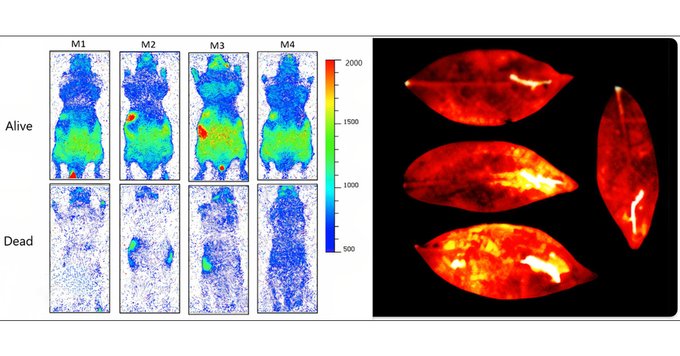
In late 2024 a University of Calgary team led by physicist Vahid Salari captured the first time-lapse images of living mice quietly flickering in total darkness and then fading to black within an hour of euthanasia. They repeated the feat with plant leaves subjected to chemical stress, watching hotspots blaze and cool as the tissues recovered. Their study, Imaging Ultra-Weak Photon Emission from Living and Dead Mice and from Plants under Stress, joins a lineage of controversial yet compelling research that suggests every living cell is, quite literally, lit from within BioRxivPubMed.
Why should readers care? Because these ghost-level photons appear to encode information about oxidative stress, metabolism, and perhaps even the cell-to-cell “conversations” that keep an organism alive. If we can learn to read this light, we may gain a new, non-invasive way to diagnose disease, monitor crops, or test environmental toxins long before classical symptoms surface. This article expands Salari’s findings into a panoramic view of UPE science—its history, mechanisms, technologies, unresolved puzzles, and real-world potential.
A Brief History of Biological Light
Early Glimmers: From Gurwitsch to Popp
-
1920s—Alexander Gurwitsch reports “mitogenetic radiation,” claiming onion roots emit ultraviolet light that stimulates cell division. The methods were rudimentary, and skepticism reigned.
-
1970s—Fritz-Albert Popp revives the idea, coining biophotons. Using photomultiplier tubes, his group records faint emissions from plant seeds, human skin, even tumor cells. He proposes that DNA may act as a coherent light source, organizing cellular processes. Critics argue instrumentation artifacts.
-
1980s-2000s bring incremental confirmation: biological tissues emit photons across 200–800 nm during oxidative reactions, but experiments are noisy, detectors imperfect, and results hard to reproduce consistently. A review by Frontiers in Physiology (2024) charts this checkered journey Frontiers.
Why the Controversy Lingered
-
Signal-to-Noise Hell – UPE intensity sits perilously close to detector dark counts.
-
Thermal Crosstalk – Living bodies also emit infrared (10⁸-fold stronger). Even minute sensor warmth can swamp optical photons.
-
The “Aura” Problem – New-age literature co-opted biophotons to justify unproven healing practices, tainting serious research by association.
Salari’s group attacked the first two obstacles with next-generation electron-multiplying CCDs (EMCCDs) cooled to -95 °C, capable of <0.002 e⁻ read noise per pixel. They neutralized the third by publishing raw spectra and kinetics, not metaphysics.
What Exactly Is Ultra-Weak Photon Emission?
Chemistry in the Dark
At its core, UPE is a form of chemiluminescence: molecules in excited electronic states relax by emitting photons. Key players include:
| Reaction Step | Molecular Actors | Photon λ (nm) |
|---|---|---|
| Lipid peroxidation | Reactive oxygen species (•O₂⁻, ¹O₂), polyunsaturated fatty acids | 500–720 |
| Protein oxidation | ROS, aromatic amino acids | 420–600 |
| DNA oxidation | ROS, nucleic bases | 350–460 |
The consensus mechanism—oxidative stress → electronically excited carbonyls or singlet oxygen → photon release—is detailed in recent reviews FrontiersNature. Unlike firefly luciferase, these reactions lack an enzyme dedicated to bright light; hence the emissions are “ultra-weak.”
Why Life Glows Only While Alive
Metabolism sustains ATP and trans-membrane gradients that feed mitochondrial electron flow, a notorious ROS generator. When circulation stops, oxygen dwindles, ROS plummet, and photon production collapses—precisely what the Calgary images record.
The Calgary Experiment: Watching Life Switch Off
Methods at a Glance
-
Subjects: 4 male C57BL/6 mice, anesthetized, maintained at 37 °C.
-
Imaging: Princeton Instruments ProEM-HS EMCCD, 30-min dark adaptation, 5-min exposures over 3 h.
-
Protocol: Baseline imaging → intraperitoneal barbiturate overdose → continued imaging.
Key Findings
-
Baseline Glow: ~1.2 × 10³ photons · cm⁻² · s⁻¹ averaged over torso—well above camera dark rate.
-
Post-mortem Decay: Intensity halved at 22 min, near baseline after 55 min, indistinguishable from system noise by 78 min.
-
Spectral Shift: Peak emission narrowed toward 560 nm during decay, implicating dwindling lipid oxidation.
A parallel plant assay using maple and oak leaves dosed with benzoin (a ROS-triggering anesthetic) displayed spatial hotspots that cooled within 16 h, confirming stress-response coupling PubMedBioRxiv.
Reading Stress in Photons—Why It Matters
Biomedical Horizons
-
Oncology: Tumor cells show elevated UPE due to chronic oxidative metabolism. Pilot rat studies of Alzheimer’s hippocampi already register disease-specific photon signatures Cell.
-
Organ Transplantation: Real-time viability assessments during perfusion could prevent graft failure.
-
Drug Toxicology: ROS-responsive chemiluminescence offers a label-free assay for mitochondrial poisons.
Agriculture & Ecology
-
Drought Monitoring – Leaves intensify UPE hours before wilting is visible.
-
Pesticide Screening – Differential photon maps reveal phytotoxic hotspots.
-
Climate Stress Biology – Correlating UPE with extreme temperature events aids crop-breeding for resilience.
Space and Astrobiology (Speculative)
If microbes under Martian regolith emit UPE when stressed by perchlorates or radiation, ultra-sensitive photometers on rovers could detect life signatures without destructive sampling.
The Technology Behind Seeing the Unseeable
Detectors
-
Photomultiplier Tubes (PMTs) – Early workhorses; superb gain but no imaging.
-
Electron-Multiplying CCDs – Photon counting with megapixel resolution; rugged, but require deep cooling.
-
sCMOS + On-chip Gain – Emerging compromise with larger fields of view at lower cost.
Noise Mitigation Strategies
-
Vacuum-sealed cryogenic housings
-
Constellations of magnetic shielding to block cosmic-ray–induced scintillation
-
Sub-nanowatt LED “dark frames” for real-time baseline subtraction
Toward Wearable “Bio-Photometers”
A 2025 Lippincott review outlines quantum dot photodiodes integrated into flexible patches that might, within a decade, map oxidative stress on human skin during athletic performance or spaceflight Lippincott Journals.
Separating Science from Pseudoscience
| Claim | Reality | Verdict |
|---|---|---|
| Biophotons prove the Kirlian “aura.” | UPE intensity is orders of magnitude below Kirlian corona discharge; the latter is driven by high-voltage ionization, not metabolism. | ❌ |
| Meditation boosts UPE coherence, enabling “healing hands.” | Small studies report lowered ROS (hence reduced UPE) after mindfulness. No evidence of external healing photons. | 🟡 Hypothesis weak |
| UPE forms a cell-to-cell optical internet. | Photonic reabsorption length in tissue ≈ 10 µm; signaling plausible only across adjacent cells or via waveguides (e.g., microtubules). | 🟠 Research needed |
Skepticism is healthy. But one cannot dismiss all UPE research as fringe—Salari’s controlled methodology and convergent evidence from plant biology anchor the phenomenon in mainstream redox biochemistry.
Open Questions That Keep Physicists Awake
-
Quantum Coherence – Do cells exploit phase relationships in emitted photons, or is the light purely stochastic?
-
Source Hierarchy – What fraction stems from mitochondria versus peroxisomes or membrane NADPH oxidases?
-
Spectral Fingerprints – Can specific ROS (superoxide, singlet oxygen, hydroxyl radical) be distinguished by their photon spectra?
-
Functional Role – Signal, by-product, or evolutionary relic? Some propose local photonic feedback regulating circadian genes; data are scarce.
-
Impact of EMF Environments – Does external electromagnetic noise modulate UPE, and could that couple to the oxidative pathways at the heart of RF-induced stress? A fertile field for cross-disciplinary inquiry.
Future Directions and Roadmap
Bench to Bedside
-
Phase I: Validate spectral biomarkers for known oxidative diseases (diabetes, Parkinson’s).
-
Phase II: Develop fiber-optic endoscopes with EMCCD tips for in vivo organ mapping.
-
Phase III: Integrate AI pattern recognition to predict pathology from photon “signatures” in real time.
Precision Agriculture
-
Drone-mounted cooled sCMOS rigs could fly night sorties, scanning hectares of crops for stress flares invisible to thermal or multispectral daylight imagery.
Regulatory and Ethical Considerations
-
Establish noise-floor standards (ISO-UPE-1) to prevent false positives.
-
Draft animal-use guidelines limiting lethal endpoints when photon decay curves suffice.
Conclusion – A Call to Look, Really Look, into the Dark
For a century biology textbooks taught that life’s business is chemical: enzymes, ions, metabolites. Salari’s mouse movies and decades of underappreciated work now insist that life is optically active as well—a candle so faint we needed exquisite detectors to notice.
Grasping UPEs is more than academic curiosity. Every photon carries a dispatch from the cellular front lines, reporting oxidative battles that precede inflammation, degeneration, or death. Learning to intercept those messages could revolutionize medicine and agronomy, revealing trouble while intervention is still easy.
As citizens, researchers, or policymakers, we stand at a threshold: either ignore this subtler spectrum or invest in technologies and studies that turn biological night-vision into tomorrow’s diagnostic gold standard. The choice, as always, is illuminated by the evidence—however weak the light may seem.
Stay curious, stay wonderful, and keep watching the darkness. It is brighter than we ever imagined.


