
The carrier tells regulators where the signal sits. The frame rate tells biology how often it returns.
Most discussions of wireless exposure begin and end with the carrier frequency: 1.9 GHz for a DECT cordless phone, 2.4 or 5 GHz for Wi‑Fi, 700 MHz or 3.6 GHz for one 5G deployment. That is the language of spectrum allocation, antenna engineering, and thermal dosimetry.
It is not a complete description of the signal a living cell encounters.
Digital wireless transmitters do not simply produce an unbroken microwave carrier. They organize transmissions in time. They burst, pause, repeat, schedule, and vary. A high-frequency carrier therefore arrives inside a lower-frequency temporal structure: frames, half-frames, time slots, beacons, duty cycles, amplitude envelopes, and traffic-dependent bursts.
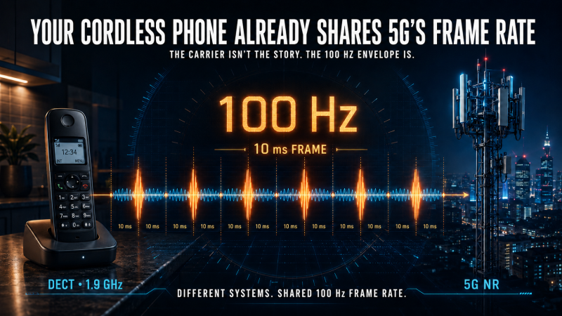
This is why an old cordless phone belongs in a modern 5G discussion. DECT and 5G are not the same technology, do not use the same carrier, and do not create identical exposure. But both are organized around a 10-millisecond radio frame. Ten milliseconds means 100 frames per second: a 100 Hz frame rate.
That shared timing does not prove a shared biological outcome. It establishes something regulators have failed to test: different wireless systems repeatedly place energy into a low-frequency timing domain that overlaps the frequencies at which living systems organize electrical and calcium-dependent work.
The public has been trained to ask, “Which gigahertz band is it?” The biologically serious question is broader:
What timing pattern is being imposed, which cellular structures can transduce it, and what happens to biological signaling fidelity when several such patterns operate together for years?
That is the question behind RF Safe’s S4–Mito–Spin and low-fidelity biology framework.
The claim, stated precisely
This article does not claim that DECT is 5G, that every 100 Hz signal is equivalent, or that one frame rate determines one disease.
It makes four narrower—and more consequential—claims:
- Modern wireless exposures have biologically relevant dimensions beyond carrier frequency and time-averaged power.
- DECT and 5G NR share a 10 ms frame architecture, while Wi‑Fi and GSM introduce other recurring low-frequency timing structures into the same environment.
- Cells use calcium oscillations, membrane voltage, mitochondrial redox state, and gene-regulatory timing as information-bearing signals; a 2026 Cell study has now identified CYB5B through a genome-wide knockout screen as essential to a 60 Hz EMF-responsive calcium-oscillation pathway.
- Current public limits do not test whether real, pulsed and modulated wireless waveforms degrade the fidelity of those biological signals under chronic, mixed-source exposure.
The last point is not rhetorical. In 2021, the U.S. Court of Appeals for the D.C. Circuit held that the FCC had failed to give a reasoned explanation for dismissing evidence concerning non-cancer effects, children, long-term exposure, pulsation or modulation, technological change, and environmental effects. The court called key parts of the agency’s decision arbitrary and capricious. The unresolved measurement problem sits in the center of that remand. Read the court’s opinion.
First, stop confusing the carrier with the temporal envelope
A simplified modulated signal can be written as:
[
E(t)=A(t)\cos\bigl(2\pi f_c t+\phi(t)\bigr)
]
Here:
- (f_c) is the radiofrequency carrier.
- (A(t)) is the changing amplitude or burst envelope.
- (\phi(t)) represents phase structure.
- The frame, slot, beacon, and traffic scheduler determine when energy is present and how it changes over time.
A spectrum regulator concentrates on (f_c). A heating standard averages the energy represented by the signal over time. But a nonlinear detector does not necessarily respond only to the average. It may respond to peaks, transitions, repetition, duty cycle, phase, or the intervals between bursts.
Biology is full of nonlinear detectors. Ion channels switch between states. Enzymes cross activation thresholds. Calcium pulses are decoded differently depending on frequency, duration, shape, baseline, and subcellular location. Mitochondria integrate local calcium microdomains and turn them into changes in ATP production, redox signaling, and reactive oxygen species.
The point is not that a 100 Hz “photon” separates from a 1.9 GHz carrier and strikes a cell. That is the wrong picture. The point is that the delivered electromagnetic field has a time structure, and biological systems may rectify, integrate, or otherwise respond to that structure.
Decades ago, Blackman and colleagues reported that 147 MHz radiofrequency radiation amplitude-modulated at low frequencies altered calcium-ion efflux from brain tissue, with responses depending on modulation frequency and power-density windows. That early result did not settle the mechanism, but it demonstrated why “carrier frequency plus average power” was already an incomplete experimental description. Blackman et al., 1980.
The household timing map
Wi‑Fi: approximately 9.77 Hz before ordinary traffic is counted
A common Wi‑Fi beacon interval is 100 time units. One Wi‑Fi time unit is 1,024 microseconds, so 100 time units equal 102.4 milliseconds. That corresponds to:
[
1/0.1024\ \text{s}=9.765625\ \text{Hz}
]
That beacon cadence is only one layer. Multiple service set identifiers, data packets, acknowledgments, retransmissions, power-saving functions, and competing devices add faster and less regular burst patterns. The result is not a pure 9.77 Hz sine wave. It is a microwave signal whose temporal structure includes a roughly 10 Hz scheduled recurrence plus traffic-dependent variability. Cisco’s explanation of the 100-TU beacon interval.
The 2026 CYB5B experiment: 60 Hz
Kim and colleagues used a controlled extremely-low-frequency magnetic field to build a remotely activated gene switch. A genome-wide CRISPR-Cas9 knockout screen identified cytochrome b5 type B, or CYB5B, as an essential mediator likely acting as an EMF sensor. Most importantly, activation depended on a distinctive pattern of rhythmic calcium oscillations, not merely on a generic rise in calcium.
That distinction is foundational. The cell responded to a calcium waveform—to timing—not simply to calcium quantity.
The exposure used in the main system was a controlled 60 Hz magnetic field at millitesla-scale flux density. It was not a Wi‑Fi router, a DECT base, or a 5G antenna. The study therefore does not establish that everyday wireless signals activate CYB5B in the same way.
But it does destroy the categorical claim that a non-heating electromagnetic field cannot be transduced into a precisely timed calcium signal and downstream gene regulation. It supplies a genetically identified mediator and a testable pathway. The proper scientific response is not to draw a “huge gap” between 60 Hz and gigahertz carrier labels and declare the question closed. It is to test whether the low-frequency envelopes, pulse trains, and switching structures carried by everyday wireless systems engage CYB5B-dependent signaling under realistic conditions. Kim et al., Cell, 2026; PubMed record.
DECT cordless phones: 100 Hz frame rate
DECT divides time into frames containing 24 full slots. Each frame lasts 10 milliseconds. The European Telecommunications Standards Institute explicitly describes the synchronization signal as 100 Hz and says it establishes the 10 ms DECT frame interval. ETSI DECT physical-layer specification.
That is not an interpretation. It is the architecture:
- 10 ms per frame
- 100 frames per second
- 24 time slots per frame
- a 100 Hz fundamental recurrence, with harmonics and device-specific pulse structure
A cordless base on a desk, nightstand, kitchen counter, nursery shelf, or office workstation is therefore not merely “a 1.9 GHz source.” It is a nearby, time-structured transmitter whose frame repeats 100 times per second.
5G NR: the same 100 Hz frame rate, plus a 200 Hz half-frame structure
The 5G New Radio physical-layer specification defines a 10 ms radio frame containing ten 1 ms subframes. Each frame is divided into two equal 5 ms half-frames. 3GPP TS 38.211 through ETSI.
That produces structural timing at:
- 100 Hz for the 10 ms frame
- 200 Hz for the 5 ms half-frame
- faster slot and symbol timing determined by numerology
- additional traffic-, beam-, and TDD-dependent variability
This does not mean every 5G exposure will show a dominant 100 Hz or 200 Hz spectral line at every location and moment. Real emissions depend on network configuration, traffic, scheduling, duplexing, beam behavior, and measurement bandwidth. It means 100 Hz and 200 Hz are built into the system’s temporal grammar and must be measured rather than erased by averaging.
GSM: approximately 217 Hz
A GSM TDMA frame lasts 120/26 milliseconds, or approximately 4.615 ms. That equals about 216.7 frames per second—the familiar nominal 217 Hz recurrence. ETSI GSM frame specification.
The important pattern
The timing map is therefore not a single frequency. It is a cluster of engineered recurrences in the low-frequency domain:
- Wi‑Fi beacon scheduling: approximately 9.77 Hz
- CYB5B gene-switch experiment: 60 Hz
- DECT frame: 100 Hz
- 5G NR frame: 100 Hz
- 5G NR half-frame: 200 Hz
- GSM frame: approximately 217 Hz
These signals differ in field type, carrier, intensity, polarization, duty cycle, waveform, source geometry, and exposure pattern. Frequency alone never makes them biologically equivalent.
But the list makes one fact unavoidable: the temporal gap is not the same as the carrier-frequency gap. Everyday wireless systems repeatedly place structured energy into the hertz-range timing domain in which biological signaling already operates.
The “huge gap” argument measures the wrong axis
The most common dismissal of the CYB5B result is visually compelling and biologically incomplete: place 60 Hz on the far left of a frequency chart, place Wi‑Fi, Bluetooth, and cellular carriers millions or billions of hertz to the right, and emphasize the empty space between them.
That chart is accurate only for the carrier axis. It omits the time-domain structure of the signal.
If the biological detector responds exclusively to carrier frequency, the gap is decisive. If it responds to envelope, switching, duty cycle, low-frequency magnetic components from device electronics, rectified fields, or repeated onset and offset, the chart has discarded the relevant variable before the experiment begins.
The correct comparison requires at least five dimensions:
- Carrier frequency
- Low-frequency envelope or frame rate
- Peak field during a burst
- Duty cycle and burst duration
- Waveform variability, polarization, and source geometry
It also requires biological measurements that preserve time:
- Calcium frequency, amplitude, phase, width, baseline, and jitter
- Membrane-potential stability and recovery
- ER calcium release and reuptake
- Mitochondrial calcium uptake, membrane potential, ATP reserve, and redox oscillations
- ROS production and antioxidant recovery
- Transcriptional and DNA-repair responses during and after exposure
Average power alone cannot answer those questions.
Calcium is not merely a mineral. It is a code.
Calcium signaling governs contraction, secretion, fertilization, immune activation, metabolism, differentiation, synaptic plasticity, transcription, apoptosis, and development. Cells distinguish signals not merely by “more calcium” or “less calcium,” but by calcium dynamics:
- Frequency: how often a pulse recurs
- Amplitude: how high the pulse rises
- Duration: how long it remains elevated
- Phase: how it aligns with other cellular oscillators
- Localization: whether it occurs near the plasma membrane, ER, nucleus, or mitochondria
- Baseline: whether calcium fully returns before the next event
- Recovery: whether pumps and stores restore the system’s prior state
A detailed review of calcium oscillations notes that downstream responses depend on frequency and on the duration, shape, baseline, and sustainability of the waveform. It also describes the ER, extracellular calcium, and mitochondria as active participants in tuning those oscillations. Dupont et al., 2016.
This matters because a cell can maintain nearly the same average calcium concentration while losing the informational fidelity of the waveform. Two signals can have the same average and opposite biological meaning.
That is the central error in a heat-only regulatory paradigm: it assumes biological significance follows average energy, while living systems often encode meaning in structure and timing.
Two possible electromagnetic front ends, one convergent calcium-redox network
RF Safe’s framework does not require every electromagnetic interaction to pass through one universal receptor. It proposes converging transduction branches whose effects are gated by tissue composition, genotype, developmental state, and recovery capacity.
Front end one: the S4 voltage sensor
Voltage-gated ion channels contain positively charged S4 helices that detect changes in membrane voltage and control channel opening. Calcium channels are among the most consequential because calcium is both charge and information.
The ion-forced-oscillation/VGIC model developed by Panagopoulos and colleagues proposes that polarized, coherent, slow-varying fields force nearby mobile ions to oscillate. Because those ions sit extremely close to S4 voltage sensors, their local Coulomb forces could perturb channel gating even when the applied field is far too weak to heat tissue appreciably.
This is a proposed biophysical mechanism, not a completed consensus mechanism. Its importance is that it is structural, quantitative, and falsifiable. It predicts dependence on frequency, polarization, intensity, ion properties, channel geometry, and waveform. It can be tested with S4 mutations, channel-specific pharmacology, patch-clamp measurements, and waveform-defined exposures. Panagopoulos et al., 2025.
Front end two: CYB5B at the mitochondrial outer membrane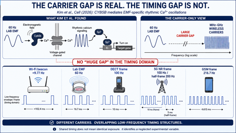
CYB5B is a membrane-bound heme protein associated with the outer mitochondrial membrane. The 2026 Cell study did not merely nominate it from a pathway diagram; a genome-wide CRISPR-Cas9 knockout screen found it essential to the EMF-responsive gene-switch mechanism. The researchers reported CYB5B-mediated, EMF-specific rhythmic calcium oscillations.
That is a major advance because it links an electromagnetic input to a genetically identified cellular component, a defined calcium pattern, and a downstream gene-regulatory result.
The exact coupling between CYB5B, calcium entry, ER calcium release, and mitochondrial calcium handling remains to be mapped. That missing bridge is now an experimental target, not a reason to return to biological denial.
Why the endoplasmic reticulum belongs in the middle
The plasma membrane and the mitochondrion are not isolated endpoints. The ER is the cell’s major intracellular calcium reservoir. IP3 receptors, ryanodine receptors, SERCA pumps, store-operated calcium entry, and mitochondria-associated membranes cooperate to generate and terminate calcium oscillations.
Mitochondria positioned close to ER calcium-release sites sample high local calcium concentrations. In doing so, they influence ATP production, metabolite flow, apoptosis, redox signaling, and the shape of the next cytosolic calcium pulse.
This produces a coupled system:
- S4-containing channels regulate calcium entry across the plasma membrane.
- ER stores amplify, pattern, and recycle intracellular calcium.
- Mitochondria decode local calcium into energetic and redox responses.
- CYB5B provides a newly identified EMF-responsive, outer-mitochondrial mediator in the 60 Hz gene-switch system.
- ROS, ATP, membrane potential, and transcription feed back into channel behavior and cellular recovery.
The biologically relevant object is therefore not one channel or one protein. It is a timed, coupled calcium–ER–mitochondrial–redox network.
S4–Mito–Spin: RF Safe’s working synthesis
RF Safe uses S4–Mito–Spin as a research framework that joins three plausible or demonstrated transduction domains:
- S4: voltage-sensor gating at the plasma membrane, especially in voltage-gated calcium and other ion channels.
- Mito: mitochondrial and ER–mitochondrial calcium handling, energetic reserve, redox signaling, and CYB5B-mediated EMF responsiveness.
- Spin: heme, flavin, iron-sulfur, and radical-pair chemistry capable of changing reaction probabilities and redox state under electromagnetic influence.
These are not interchangeable mechanisms. They are possible parallel entry points into the same information-processing network.
RF Safe adds a persistence gate: cell longevity, turnover, repair, antioxidant reserve, immune surveillance, and exposure recovery. A transient signal in a rapidly renewed, well-buffered tissue may be erased. The same signal in a long-lived, metabolically active, poorly recovering cell lineage may persist, accumulate, or alter later decisions.
This is density gating:
- Tissues differ in ion-channel abundance and excitability.
- Cells differ in mitochondrial density and metabolic throughput.
- Redox-active proteins and cofactors differ across lineages.
- Genotypes alter channel expression and response thresholds.
- Developmental windows alter both sensitivity and consequence.
- Long-lived cells retain history that high-turnover tissues can dilute.
The model therefore predicts heterogeneity—not uniformity. It predicts effects in some tissues, exposure windows, waveforms, and genotypes, with null results in others. It predicts nonlinear windows rather than a simple “more power equals more effect” curve.
That pattern is often treated as a weakness in the non-thermal evidence. In a timing- and density-gated system, it is exactly what should be expected.
Human genotype already shows that the “receiver” matters
In a 2025 double-blind, sham-controlled experiment, 34 healthy participants were exposed before sleep to standardized 5G signals at 3.6 GHz and 700 MHz. Researchers tested a common non-coding variant in CACNA1C, the gene encoding the principal pore-forming subunit of the Cav1.2 L-type calcium channel.
The 3.6 GHz exposure shifted sleep-spindle center frequency in T/C carriers of rs7304986 but not in matched T/T carriers. The effect appeared across central, parietal, and occipital regions. Sousouri et al., NeuroImage, 2025.
The point is not that this single study proves harm. The point is that a controlled RF exposure produced an objective physiological response that depended on common genetic variation in a voltage-gated calcium-channel gene.
Same protocol. Different receiver.
Population averages can therefore dilute a real subgroup response. A safety standard that models “the public” as a thermally uniform mass misses biologically meaningful variance at the channel, mitochondrial, genomic, and developmental levels.
From timing noise to oxidative stress
Reactive oxygen species are not merely damage molecules. At controlled levels they are signaling intermediates. They help regulate adaptation, immunity, differentiation, mitochondrial turnover, and repair. The problem is not the existence of ROS; it is loss of redox control—too much production, mistimed production, inadequate antioxidant buffering, or failure to return to baseline.
A calcium-timing disturbance can raise oxidative burden through several routes:
- Excess or mistimed mitochondrial calcium can destabilize electron transport.
- Ion-channel dysfunction can increase energetic demand on pumps that restore gradients.
- NADPH oxidases and nitric-oxide synthases can shift ROS and reactive-nitrogen output.
- ER stress and mitochondrial stress can reinforce one another.
- Persistent redox disturbance can alter proteins, lipids, DNA, chromatin, transcription, and repair signaling.
The oxidative-stress literature is too large to dismiss as isolated noise. A 2016 review found oxidative effects in 93 of 100 peer-reviewed low-intensity RFR studies available to its authors, including ROS-generating pathways, lipid peroxidation, oxidative DNA damage, and altered antioxidant enzymes. Yakymenko et al., 2016.
Henry Lai’s continuously updated literature compilations report the same broad direction across a much larger body of work. As of the June 2026 update summarized by ICBE-EMF, 390 of 438 RFR oxidative/free-radical papers reported significant effects, including 110 of 114 studies at SAR values at or below 0.40 W/kg. The compilations also report majorities of effect findings in genetic, neurological, gene-expression, reproductive, and developmental research. Lai compilation and downloadable abstracts; ICBE-EMF summary.
These counts are not a formal meta-analysis and do not calculate population disease risk. They answer a more basic question: is biological interaction below heating thresholds an occasional anomaly, or is it a recurrent experimental signal? The answer is recurrent.
The U.S. National Toxicology Program adds controlled evidence at organism scale. Under its study conditions, the NTP found clear evidence of malignant heart schwannomas in male rats, some evidence of malignant brain gliomas, and RFR-associated DNA damage in specific brain regions and blood cells. NTP explicitly states that the mechanism of the DNA damage was not determined. NTP radiofrequency research summary.
That unresolved mechanism is precisely where calcium timing, S4 gating, mitochondrial redox, and CYB5B now belong.
The household is a mixture, not a single-source chamber
Real indoor exposure is rarely one clean waveform.
A home may contain:
- A DECT base repeating on a 100 Hz frame
- A Wi‑Fi access point emitting scheduled beacons and traffic bursts
- Phones performing network registration, data transfer, and background synchronization
- Bluetooth wearables and peripherals
- Smart meters, monitors, cameras, appliances, and neighboring networks
- Low-frequency magnetic and electric fields from wiring and power electronics
The organism encounters the sum in time, not a regulatory spreadsheet that evaluates each source separately.
This matters because coupled biological oscillators can respond to repetition, beat patterns, phase relationships, recovery intervals, and stochastic variability. A second source need not add enough energy to cause heating. It may add timing noise before the system has recovered from the first source.
A 2023 lettuce study is useful because it exposed plants to a realistic mixture of ordinary DECT and dual-band Wi‑Fi equipment. Greenhouse effects were limited, but field-grown plants showed reduced photosynthetic efficiency, earlier flowering, down-regulation of VDE and ZEP genes involved in photoprotection, and impaired handling of light stress. The experiment did not include separate DECT-only, Wi‑Fi-only, continuous-wave, and 100 Hz pulsed arms, so it cannot identify the causal waveform. Its value is different: an ordinary mixed indoor wireless environment was biologically active enough to alter stress handling under some growth conditions. Tran et al., Plants, 2023.
The correct response is not to pretend that study proved “100 Hz caused the effect.” It is to perform the missing factorial experiment.
Low-fidelity biology: the upstream problem
RF Safe uses low-fidelity biology to describe a state in which the timing, spatial precision, recovery, and error-correction capacity of biological signaling are degraded.
The initiating disturbance is bioelectrical dissonance: an imposed environmental signal that perturbs the electrical and redox rhythms by which cells coordinate metabolism, repair, development, immunity, and collective behavior.
This is not a claim that RF exposure directly causes a list of unrelated diseases. It is a meta-disease framework:
- A time-structured electromagnetic exposure interacts with one or more transduction pathways.
- Membrane gating, calcium signaling, ER release, mitochondrial handling, or redox chemistry becomes less precise.
- ATP reserve, antioxidant recovery, transcription, and repair carry a higher background load.
- The organism compensates, but compensation consumes reserve and may shift gene expression or cell state.
- Tissue density, genotype, age, development, co-exposures, and cell longevity determine where the disturbance persists.
- Downstream outcomes emerge opportunistically rather than as one inevitable disease.
In this model, the signal does not need to “cause cancer,” “cause infertility,” or “cause a neurological disorder” in a one-step chain. It needs only to reduce the fidelity of the upstream systems that normally keep rare errors rare, defer age-related failure, terminate inflammation, repair DNA, remove damaged cells, and restore metabolic baseline.
The signature predictions are therefore:
- Rare outcomes become less rare.
- Later-life failures appear earlier.
- Recovery takes longer.
- A second stressor has a larger effect because reserve is already reduced.
- Developmental exposures have disproportionate consequences because timing helps construct the system itself.
- Different tissues produce different endpoints because their channels, mitochondria, redox hardware, and persistence gates differ.
- Beneficial, adverse, mixed, nonlinear, and null results can all occur because an upstream control system is being perturbed rather than a single toxin acting through a single dose-response pathway.
This is why “the epidemiology does not point cleanly to one disease” is not a rebuttal to the framework. A loss of computational fidelity should widen the distribution of downstream outcomes, not collapse it into one diagnosis.
Why thermal compliance cannot certify biological fidelity
Current FCC and ICNIRP limits are organized around absorbed power, temperature rise, and time averaging. Those variables matter for preventing excessive heating. They are not designed to bound every non-thermal interaction with an electrically excitable, rhythm-dependent organism.
Time averaging can make two exposures appear equal even when their peak structure is radically different. A continuous exposure and a train of intense, short bursts can share the same average power while presenting different onset, recovery, and repetition patterns to a nonlinear biological system.
The regulatory blind spots include:
- Frame rate and low-frequency envelope
- Pulse width and peak-to-average ratio
- Duty cycle and recovery interval
- Phase, polarization, and waveform variability
- Multiple simultaneous sources
- Chronic exposure beginning before birth
- Genotype and developmental susceptibility
- Tissue-specific channel and mitochondrial density
- Calcium waveform fidelity rather than calcium quantity
- Redox recovery, repair, and persistence after exposure ends
ICBE-EMF’s peer-reviewed assessment states directly that limits based on time-averaged SAR or power density, without considering amplitude or frequency modulation, do not adequately address potential effects of real-world RFR. ICBE-EMF analysis in Environmental Health, 2022.
The D.C. Circuit did not order the FCC to declare that one wireless technology causes one disease. It ordered the agency to provide a reasoned explanation. A reasoned explanation in 2026 must address the variables that the new science can now name and test.
The decisive experiment regulators should fund now
The scientific dispute can be made far more tractable. Build an exposure matrix that separates carrier, envelope, peak, duty cycle, and field type while measuring the biological signal in real time.
Exposure arms
- Sham exposure
- Pure 60 Hz ELF magnetic-field replication of the Kim protocol
- Pure 100 Hz and 200 Hz ELF conditions
- Continuous-wave RF carriers without low-frequency modulation
- The same carriers amplitude-modulated or pulse-gated at 9.77, 60, 100, 200, and 216.7 Hz
- Recorded and faithfully reproduced DECT, Wi‑Fi, GSM, LTE, and 5G waveforms
- DECT plus Wi‑Fi and other defined mixtures
- Equal-average-power, different-peak-and-duty-cycle comparisons
- Acute, repeated, chronic, and post-exposure recovery protocols
Mechanistic interventions
- CYB5B knockout, rescue, and controlled overexpression
- S4 voltage-sensor mutations and channel-specific blockers
- L-type and T-type calcium-channel interventions
- ER store depletion and selective IP3R/RyR manipulation
- Mitochondrial calcium uniporter and VDAC manipulation
- Radical scavengers and redox-pathway interventions
High-fidelity readouts
- Calcium frequency, amplitude, phase, jitter, localization, and termination
- Patch-clamp measurements of channel gating
- Membrane potential and recovery time
- ER calcium release and refilling
- Mitochondrial membrane potential, calcium, ATP, NADH/NAD+, and oxygen consumption
- ROS location, pulse timing, antioxidant reserve, and post-exposure recovery
- Chromatin accessibility, transcription, DNA damage, and repair kinetics
- Senescence, apoptosis, immune signaling, differentiation, and cell-state transitions
- Single-cell and spatial analyses instead of population averages alone
Susceptibility variables
- CACNA1C and other channel-related genotypes
- Differentiation state
- Mitochondrial and channel density
- Developing versus mature tissue
- Short-lived versus long-lived cell lineages
- Normal versus metabolically stressed conditions
If CYB5B is irrelevant to telecom-like envelopes, knockout and rescue studies will show it. If S4 is irrelevant, voltage-sensor manipulation will show it. If 100 Hz frame structure is biologically indistinguishable from continuous RF at equal average power, calcium imaging and electrophysiology will show it.
But until those experiments are done, carrier-frequency separation is not an answer. It is a reason to perform the correct experiment.
What families can do now
The policy failure is collective, but exposure reduction does not require abandoning communication.
- Replace DECT cordless phones with corded phones where practical.
- If DECT is retained, use a verified low-radiation/Eco mode and keep the base away from beds, nurseries, desks, and other long-occupancy locations.
- Turn off unnecessary wireless transmitters during sleep.
- Use Ethernet for fixed computers, televisions, game systems, and access points.
- Keep actively transmitting phones away from the body when a wired or speaker option is available.
- Prefer wired and optical networking—especially in schools, childcare settings, hospitals, and bedrooms.
- Make Li‑Fi compatibility a standard feature so indoor connectivity can increasingly move from pulsed microwaves to light-based links.
These are engineering choices, not a rejection of technology. The goal is biocompatible connectivity: move high-bandwidth indoor data into wires and light, and reserve radiofrequency transmission for the places where mobility genuinely requires it.
The policy conclusion: regulate the signal biology actually receives
DECT’s 1.9 GHz carrier is not 5G. Its 100 Hz frame rate, however, is the same frame rate built into 5G NR’s 10 ms radio frame. Wi‑Fi adds a roughly 9.77 Hz beacon cadence. GSM adds a roughly 217 Hz frame recurrence. Real traffic adds variability, bursts, and incomplete recovery intervals.
At the same time, modern biology shows that:
- Calcium oscillations carry information in their timing and shape.
- The ER and mitochondria actively construct and decode those waveforms.
- S4 voltage sensors provide a falsifiable membrane-level transduction hypothesis.
- A genome-wide knockout screen has identified CYB5B as essential to a 60 Hz EMF-responsive calcium-oscillation and gene-switch pathway.
- Common CACNA1C variation can alter an objective human brain response to standardized 5G exposure.
- Oxidative, genetic, neurological, reproductive, and developmental effects recur across a large experimental literature at non-heating exposure levels.
The missing bridge is no longer mysterious. It is experimentally approachable.
The regulatory question is therefore not whether a 1.9 GHz photon and a 3.6 GHz photon are the same. They are not. The question is whether different wireless carriers can impose overlapping low-frequency timing structures on calcium- and voltage-dependent biology—and whether chronic mixtures degrade biological fidelity long before tissue heats.
That question has not been answered by SAR.
It has not been answered by averaging power for minutes.
It has not been answered by drawing a “huge gap” between 60 Hz and a microwave carrier while deleting the envelope from the graph.
And it cannot be answered by assuming that legal exposure is biologically silent.
Different systems. Shared timing domain. Unfinished science. Unprotected biology.
The next generation of exposure standards must measure the signal that biology actually receives: carrier, envelope, pulse, peak, duty cycle, mixture, timing, and recovery. Until then, thermal compliance is not proof of biological compatibility.
Core references
- Kim J, Hwang Y, Kim S, et al. Electromagnetic field-inducible in vivo gene switch for remote spatiotemporal control of gene expression. Cell. 2026;189(11):3465–3480.e23. DOI and full record.
- ETSI. 3GPP TS 38.211: NR; Physical channels and modulation. Official specification.
- ETSI. EN 300 175-2: DECT Common Interface; Physical Layer. Official specification.
- ETSI. GSM frame and time-slot structure. Official specification.
- Blackman CF, et al. Induction of calcium-ion efflux from brain tissue by radiofrequency radiation. Bioelectromagnetics. 1980;1:35–43. PubMed.
- Panagopoulos DJ, Yakymenko I, De Iuliis GN, Chrousos GP. A comprehensive mechanism of biological and health effects of anthropogenic extremely low frequency and wireless communication electromagnetic fields. Front Public Health. 2025;13:1585441. Full text.
- Sousouri G, et al. 5G radio-frequency-electromagnetic-field effects on the human sleep electroencephalogram: A randomized controlled study in CACNA1C genotyped volunteers. NeuroImage. 2025;317:121340. Open record and full text.
- Dupont G, et al. Fine tuning of cytosolic Ca2+ oscillations. F1000Research. 2016. Full text.
- Yakymenko I, et al. Oxidative mechanisms of biological activity of low-intensity radiofrequency radiation. Electromagn Biol Med. 2016;35(2):186–202. PubMed.
- National Toxicology Program. Cell Phone Radio Frequency Radiation. Official research summary.
- Environmental Health Trust v. FCC, 9 F.4th 893 (D.C. Cir. 2021). Court opinion.
- International Commission on the Biological Effects of Electromagnetic Fields. Scientific evidence invalidates health assumptions underlying the FCC and ICNIRP exposure limit determinations for radiofrequency radiation. Environmental Health. 2022;21:92. Full text.
- Tran NTA, et al. Effects of non-ionizing electromagnetic radiation on lettuce plants in field and greenhouse conditions. Plants. 2023;12(5):1082. PubMed.
- Lai H. Effects of exposure to electromagnetic fields: Thirty years of research. Abstract collections and summaries.
RF Safe presents low-fidelity biology and S4–Mito–Spin as a mechanistic research framework: a synthesis intended to generate testable predictions about upstream biological signaling, not a clinical diagnosis or a claim that one exposure produces one inevitable disease.