
S4–Mitochondrial–Spin Model
Research hypothesis and evidence review
Prepared for the RF Safe research program
Abstract
Radiofrequency electromagnetic-field (RF-EMF) experiments do not produce a uniform biological pattern. In the major positive lifetime rodent bioassays, the clearest repeated tumor signal is concentrated in glial lineages: malignant glioma in brain and malignant schwannoma in the heart. Hardell’s positive human case-control findings likewise center on glioma and acoustic neuroma, a Schwann-cell tumor. Shorter-term cellular experiments report effects in neural and glial models while many skin-cell experiments are negative for their selected endpoints. This recurring concentration of positives is evidence of tissue-selective susceptibility. Here, that pattern is treated as the phenomenon to be explained.
We formulate density gating as a falsifiable hypothesis within an S4–mitochondrial–spin framework. Density is plural and tissue dependent. In one tissue, the dominant lever may be the number and metabolic activity of mitochondria or other redox organelles. In another, it may be the surface density and state of S4-containing voltage sensors and the resulting bulk or oscillatory calcium throughput. In another, it may be the abundance of CYB5B or other heme-, flavin- or iron–sulfur-containing electron-transfer systems capable of redox and spin-sensitive transduction. These branches can operate separately or reinforce one another. Antioxidant defense, repair, adaptation and tissue turnover determine whether the resulting signal is erased or persists.
The core tissue pattern is directly observed. The US National Toxicology Program (NTP) reported exposure-related heart schwannomas and brain gliomas in male rats under chronic GSM- and CDMA-modulated exposures; the Ramazzini Institute again reported heart schwannomas under a much lower far-field exposure. Human umbilical-cord-blood cells exposed to a UMTS signal showed a transient ROS response graded by differentiation state. Primary astrocytes have shown a modulation- and duration-dependent ROS/DNA-fragmentation response, whereas controlled keratinocyte studies have often been null or weak for their selected endpoints. Independent biology establishes the density gradients that the hypothesis requires: cardiomyocyte mitochondria occupy roughly 25–30% of cell volume; mitochondria are allocated to high-demand neural and Schwann-cell microdomains; differentiating epidermal keratinocytes progressively fragment and eliminate mitochondria; and S4-channel expression and calcium coupling vary with lineage and state.
The observed clustering is evidence for density-gated susceptibility, while the relative contribution of each density lever remains to be measured directly. No pivotal RF study has yet quantified all proposed susceptibility variables across multiple cell types under identical dosimetry. A contemporary cohort reports null associations, but serious published criticisms of its exposure classification, reference group, latency, statistical power and tumor localization materially limit its value as counterevidence to the positive Hardell findings. This paper therefore explains the observed tissue pattern, defines a multi-branch susceptibility index, and proposes experiments that can determine which density lever dominates in each tissue.
Keywords: radiofrequency electromagnetic fields; tissue susceptibility; density gating; S4 voltage sensor; voltage-gated ion channel; calcium; mitochondria; reactive oxygen species; radical pair; Schwann cell; glia; differentiation
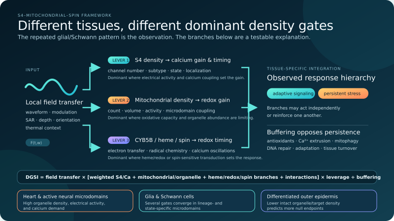
Figure 1. Different tissues can be gated by different densities. Local dosimetry supplies the input. Tissue susceptibility can then be dominated by S4/calcium density, mitochondrial/organelle density, or CYB5B/heme/redox/spin-active density, with interaction terms when the branches converge. Antioxidants, calcium extrusion, mitophagy, DNA repair and tissue turnover oppose persistence.
1. The question is not merely whether an effect exists, but where it can be amplified
If RF-EMF produced biological effects through a single uniform bulk-heating mechanism, responses should largely track absorbed energy and thermal physiology. If a reproducible nonthermal pathway also exists, uniformity is not required. Biology is full of nonlinear transducers: a small initiating perturbation can be amplified in a cell that is near a gating threshold, tightly couples calcium to mitochondria, or has limited antioxidant reserve, while the same perturbation is buffered in a neighboring cell.
This motivates the central question:
At the same adequately characterized local exposure, do cells with a greater effective density and coupling of susceptible voltage-sensing, calcium-handling, mitochondrial and radical-redox elements show a larger and more persistent response?
The phrase effective density includes literal abundance but does not stop there. Mitochondrial count or volume can be the dominant determinant where oxidative capacity is the limiting lever. S4-channel number can dominate where field-sensitive gating and calcium throughput are limiting. Heme/redox/spin-active protein abundance can dominate where electron-transfer chemistry is limiting. Localization and operating state then determine how much of that inventory is biologically available.
The proposed model predicts conditional susceptibility through multiple density gates. It does not require the same lever to dominate every tissue.
2. What the evidence establishes—and what it does not
2.1 Chronic rodent bioassays show repeated glial and Schwann-cell enrichment
The NTP exposed rats from gestation through two years to 900 MHz GSM- or CDMA-modulated fields in 10-minute on/off cycles, with whole-body specific absorption rates of 1.5, 3 or 6 W/kg. Its final report concluded that there was clear evidence of carcinogenic activity in male rats based on malignant schwannomas of the heart. Malignant gliomas and glial hyperplasias in the brain contributed additional evidence. In male rats, heart schwannomas occurred in 0/90 sham controls and 2/90, 1/90 and 5/90 animals in the GSM groups; the corresponding CDMA counts were 0/90, 2/90, 3/90 and 6/90. Brain gliomas were rarer and their dose patterns were less monotonic. Other organs produced weaker, mixed or equivocal findings.[1]
The NTP genotoxicity component was similarly heterogeneous. After subchronic exposure, comet-assay DNA damage was positive in male-rat hippocampus for CDMA, male-mouse frontal cortex for both modulations, and female-mouse leukocytes for CDMA, while numerous tissue/modulation combinations were negative or equivocal and blood micronucleus tests were negative.[2] This pattern is compatible with tissue-, sex- and waveform-dependent susceptibility, but it cannot identify the mechanism by itself.
The Ramazzini Institute exposed 2,448 rats from prenatal life until natural death to a 1.8 GHz GSM far field for 19 hours per day at 0, 5, 25 or 50 V/m. The investigators reported a statistically significant increase in heart schwannomas in males at 50 V/m, increases in Schwann-cell hyperplasia in males and females, and a nonsignificant increase in malignant glial tumors in females.[3] The recurrence of rare Schwann-cell and glial histotypes across two different exposure systems is mechanistically interesting.
A 2025 WHO-commissioned systematic review of animal cancer studies rated the evidence for increased glioma and heart schwannoma in male rats as high certainty.[4] A 2026 commentary led by Karipidis disputed that judgment on methodological and analytical grounds.[5] Its institutional position is relevant and must be disclosed: six of its seven authors were affiliated with the Australian Radiation Protection and Nuclear Safety Agency (ARPANSA), including Karipidis. He is ARPANSA’s Assistant Director of Health Impact Assessment, has served on working groups developing the Australian RF exposure standard, joined the ICNIRP Main Commission in 2020 and has served as ICNIRP Vice Chair since July 2024.[38,39] ARPANSA states both that its RF standard was aligned with the 2020 ICNIRP guidelines and that excessive heating at high exposure levels remains the only established RF-EME health effect.[40]
This creates a structural role overlap: a senior participant in the institutions that develop, apply and publicly defend the prevailing exposure-limit framework is also leading a critique of evidence that could challenge the adequacy of that framework. It does not prove that any technical argument in the commentary is wrong, and it is not a substitute for point-by-point methodological evaluation. But the commentary is not independent external validation and should not be presented as though it were. A declaration of no financial competing interest does not exhaust nonfinancial institutional commitments or conflicts of perspective.
The central observation remains: among the tumor findings, glial and Schwann-cell lineages provide the clearest repeated cross-study convergence. Density gating is proposed to explain that convergence. The unmeasured question is not whether the pattern exists, but which combination of mitochondrial, S4/calcium and heme/redox/spin density produced it.
2.2 Human evidence contains a positive lineage-specific signal and a contested cohort null
Hardell and colleagues reported associations between long-term wireless-phone use and glioma or acoustic neuroma, including stronger estimates for ipsilateral use and long latency.[6,7] This is directly aligned with the tissue-selective pattern: central glia and peripheral Schwann cells again emerge rather than a uniform distribution of tumor types.
They are not the whole human evidence base. In the prospective COSMOS cohort, 264,574 participants contributed more than 1.8 million person-years. Regression-calibrated cumulative call time was not associated with glioma, meningioma or acoustic neuroma; the glioma hazard ratio for the highest cumulative-use category was 1.07 with a wide 95% confidence interval of 0.62–1.86.[8] That is a reported null association, but it is not a precise exclusion of risk: the interval is compatible with anything from a 38% reduction to an 86% increase. For use longer than 15 years, the glioma estimate of 0.97 had a 95% confidence interval of 0.62–1.52, likewise unable to exclude a clinically meaningful increase.[8]
Moskowitz, Frank, Melnick, Hardell and colleagues published a detailed critique of COSMOS in the same journal.[36] Its most consequential points are directly relevant to density gating:
- Call time is not absorbed RF dose. COSMOS calibrated self-reported call duration with operator records from a subset, but handset output can vary by orders of magnitude with network technology, signal strength and adaptive power control. Calibration can improve the estimate of minutes while still misclassifying the biologically relevant waveform and tissue dose.
- Exposure was anchored at baseline. The tumor analysis used baseline mobile-phone histories to predict outcomes over a median 7.12-year follow-up while phones, networks, use patterns and tower density changed. Other RF sources—including cordless phones, Wi-Fi, personal wireless devices and environmental transmitters—were not incorporated into the primary dose metric.
- There was no unexposed reference group. Nearly all participants were already users, and roughly two-thirds had used mobile phones for at least ten years. COSMOS compared the highest groups with the bottom 50% of an already-exposed distribution—a reference category reported in the critique as averaging as much as 10.6 minutes of calls per day. Exposure overlap and measurement error reduce contrast and can pull estimates toward the null.
- The lineage-specific tests were underpowered. Only 149 gliomas and 29 acoustic neuromas occurred. The wide confidence intervals cannot exclude moderate or even substantial increases, especially in the longest-latency strata.
- Anatomical specificity was not used. The main analysis did not test ipsilateral versus contralateral tumors, frontal or temporal location, cumulative cordless-plus-mobile exposure, or glioblastoma separately—the very spatial and histologic distinctions needed to test a localized density-gating prediction.
- Outcome and country heterogeneity remain concerns. The critique identified possible under-ascertainment of benign tumors in registries, unexpected glioma/meningioma incidence patterns, significant UK-versus-other-country heterogeneity for some outcomes, and large country differences in what counted as the upper exposure tertile.
- Funding deserves disclosure in interpretation. Parts of COSMOS in Finland, Sweden and the United Kingdom received telecommunications-industry funding through arrangements described as scientific firewalls. This does not by itself invalidate the data, but it is a relevant competing-interest consideration when a null result is used for policy reassurance.
The COSMOS investigators replied that the prospective design avoids differential recall after diagnosis, regression calibration improves call-time estimates, non-users would be an unusually selected and confounded comparison group, and national registries provide high-quality outcome ascertainment.[37] The reply’s lead author, Maria Feychting, served as an ICNIRP Commission member from 2008 to 2020 and as its Vice Chair from 2012 to 2020.[45] That history should be disclosed when the exchange is used to inform exposure policy, while the validity of the reply must still be judged from its methods and reasoning. Its defenses of the cohort design are legitimate points. They do not, however, turn call duration into tissue-specific RF dose, create a genuinely low-exposure comparison group, lengthen follow-up, increase the number of acoustic neuromas, narrow the reported confidence intervals, or supply the missing laterality and tumor-location analyses.
A 2024 WHO-commissioned systematic review likewise concluded that mobile-phone exposure likely does not increase risk of the most studied brain tumors.[9] It would be inaccurate, however, to present that review as an institutionally detached adjudication of the ICNIRP exposure framework. Karipidis had been an ICNIRP Commissioner since 2020 and became Vice Chair in July 2024; coauthor Dan Baaken became ICNIRP Scientific Secretary and a Board member in July 2024; and coauthor Martin Röösli served as an ICNIRP Commissioner from 2016 to 2024 and remains in its Scientific Expert Group.[38,43,44] The review therefore includes substantial authorship overlap with the organization whose guidelines it can be used to validate.
Frank, Moskowitz, Melnick, Hardell and colleagues subsequently published a peer-reviewed critique alleging serious flaws in exposure classification, eligibility decisions, risk-of-bias judgments and interpretation.[41] Karipidis and colleagues published a response defending their methods and, in that response, expressly disclosed Karipidis’s governmental risk-advice role and his ICNIRP work developing and disseminating exposure-limitation advice.[42] The correct scholarly treatment is to show both sides and then assess the disputed decisions. The absence of a declared financial conflict does not eliminate the structural conflict of perspective created when evidence reviewers also hold senior roles in the guideline-setting system reinforced by their conclusions.
The review’s conclusion should therefore be reported as one contested synthesis, not used to erase limitations in its component evidence or the positive lineage-specific findings. For the present hypothesis, COSMOS is best characterized as an imprecise null under substantial exposure misclassification, not as decisive evidence against a glioma or Schwann-cell signal.
Density gating also gives a mechanistic reason population averages can dilute an effect: heterogeneous waveform and anatomical coupling are layered on genotype, cell state and a potentially small high-susceptibility subgroup. Human testing should therefore combine dosimetry, laterality, tumor lineage and susceptibility biomarkers rather than treating cumulative call minutes or all users as biologically equivalent.
2.3 Skin nulls are an expected low-density contrast
Skin receives substantial superficial RF energy, particularly as frequency rises and penetration depth falls. Yet several controlled keratinocyte experiments have been largely negative for the endpoints examined. Human keratinocytes and reconstructed epidermis exposed to GSM-900 at 2 W/kg for 48 hours showed no apoptosis, altered proliferation or thickness, or heat-shock-protein change in isolated keratinocytes; small culture-dependent heat-shock changes occurred in reconstructed epidermis and fibroblasts.[10] A reverberation-chamber study of normal human keratinocytes at 900 MHz found no reproducible gene-expression response after short, low-SAR exposures.[11]
These nulls are not surprising under density gating. Skin cells are not devoid of the relevant machinery: keratinocytes express functional CaV1.2, and channel agonists and antagonists alter epidermal barrier recovery.[12] They also respire and generate mitochondrial ROS.[13] The distinction is quantitative and spatial. Cardiomyocyte mitochondria occupy roughly 25–30% of cell volume, and mitochondria concentrate at high-demand neural and Schwann-cell microdomains.[30–33] In the epidermis, differentiating keratinocytes undergo programmed mitochondrial fragmentation and wholesale organelle clearance as they move toward the surface.[34] Thus much of the exposed outer epidermis contains a lower average density of intact mitochondria, active S4-calcium machinery and long-lived susceptible cellular material than heart and neural tissues.
The controlled skin nulls therefore strengthen rather than weaken the density-gating picture: high superficial exposure does not guarantee a positive result when the biological target density is lower. Basal keratinocytes, fibroblasts, melanocytes, immune cells and cutaneous nerves will not share one score, but the differentiated epidermis supplies the low-density comparison the hypothesis predicts.
3. Mechanistic pillar one: S4 voltage sensors as a variable biological front end
Voltage-gated sodium, calcium and potassium channels contain positively charged S4 transmembrane segments that move within the membrane electric field and control channel gating. The density, subtype, membrane localization, gating-charge structure and resting-state occupancy of these channels differ greatly by cell type and differentiation state.
Panagopoulos and colleagues’ 2025 IFO–VGIC model proposes that low-frequency modulation, pulsation and variability within wireless signals can force mobile ions near voltage-gated ion channels to oscillate and thereby exert coordinated Coulomb forces on S4 voltage sensors.[14] This is a specific biophysical proposal, not an experimentally established explanation of the NTP tumors. Its value for density gating is that it identifies a potentially countable front end: if the mechanism is correct, more appropriately localized voltage sensors operating in a responsive state should increase the probability of transduction.
Pall’s 2013 paper reviewed 23 studies in which calcium-channel blockers reduced diverse EMF-associated responses and proposed a VGCC–calcium–nitric-oxide/peroxynitrite pathway.[15] Two qualifications matter. First, Pall synthesized experiments conducted by multiple groups; the blocker experiments were not all performed by Pall. Second, the exposures included static, extremely low-frequency, pulsed and microwave fields, so their aggregation does not by itself establish a single radiofrequency mechanism. Blockers can also have off-target effects.
Direct electrophysiology supplies both support and resistance. A systematic review of neuronal ion-channel experiments found that calcium homeostasis was the most frequently reported target and that outcomes depended on frequency, duration and intrinsic channel expression.[16] In contrast, Platano and colleagues found no effect of acute low-level continuous-wave or GSM-modulated 900 MHz exposure on Ba2+ currents through voltage-gated calcium channels in rat cortical neurons.[17] Under much stronger nanosecond pulsed electric fields, cells expressing VGCCs showed roughly twice the conductance increase of VGCC-negative cells, and susceptibility correlated with pre-exposure VGCC current.[18] That nanosecond electroporation result is not evidence that ordinary wireless RF acts the same way; it is proof of the narrower biological principle that measured channel expression can stratify cellular electrical-field susceptibility.
S4 density is dynamic, not an anatomical constant
Schwann cells express neuronal-type voltage-gated sodium and potassium channels, and functional channel density changes with proliferation and myelination.[19,20] Voltage-gated calcium currents in mammalian Schwann cells can depend on coculture, development and cell state.[21] Astrocyte CaV expression likewise changes during reactive states.[22] Thus, a mature quiescent Schwann cell, a repair Schwann cell and a proliferating Schwann-cell precursor should not receive the same susceptibility score.
This state dependence offers a way to explain positive and null results without making the hypothesis unfalsifiable: channel abundance and operating state must be measured before exposure, and the resulting score must predict outcome in samples not used to fit the model.
4. Mechanistic pillar two: calcium–mitochondrial coupling as the gain stage
Mitochondria are not simply batteries. Calcium uptake regulates metabolism, and calcium overload or disturbed redox balance can increase ROS, alter membrane potential, remodel mitochondrial networks and trigger cell-death or stress pathways. The effect of an ion-channel perturbation should therefore depend on physical and biochemical coupling between the plasma membrane, endoplasmic reticulum and mitochondria—not merely on total mitochondrial mass.
This coupling is prominent in neural support cells. Electrical stimulation of peripheral axons releases ATP, activating a P2RY2–IP3–mitochondrial-calcium-uniporter pathway in myelinating Schwann cells. Suppression of this neuron-to-Schwann-cell mitochondrial calcium signal impaired myelination in vivo.[23] The result does not involve RF, but it demonstrates a high-gain, activity-linked route by which extracellular electrical activity can reach Schwann-cell mitochondria.
Primary astrocytes provide a more direct RF observation. Campisi and colleagues exposed differentiated rat astroglial cultures to either continuous 900 MHz fields or 900 MHz fields amplitude-modulated at 50 Hz. Significant ROS elevation and DNA fragmentation occurred only after 20 minutes of modulated exposure, not with shorter durations or continuous-wave exposure.[24] This single study requires replication, but its waveform and time dependence are the kind of interaction density gating predicts.
A real-time microscopy study reported that pulsed 2.856 GHz exposure altered cytosolic, endoplasmic-reticulum and mitochondrial calcium in primary hippocampal neurons at 4 and 40 W/kg, whereas primary cardiomyocytes responded only at 40 W/kg; mitochondrial calcium was the most sensitive compartment measured.[25] Because 40 W/kg is high and thermal microgradients and exposure artifacts require stringent control, the study is not evidence for everyday risk. It is nonetheless a direct example of cell-type and organelle-specific response thresholds.
Human cord blood supplies a differentiation-state experiment
Durdik and colleagues exposed human umbilical-cord-blood mononuclear cells to a 1,947.4 MHz UMTS signal at an SAR of 40 mW/kg. After one hour, ROS increased by 22% in CD45+ lymphocytes and 27% in CD34+ hematopoietic stem/progenitor cells; within CD34+ cells, the 28% increase in progenitors reached borderline significance while the increase in the least differentiated CD34+CD38− stem-cell fraction did not. No exposure effect remained at three hours. Baseline ROS followed the differentiation sequence stem cell < progenitor < lymphocyte, consistent with increasing mitochondrial metabolism. The exposure did not cause persistent DNA damage, apoptosis or preleukemic fusion genes.[26]
This is one of the most relevant observations for density gating because cell state was compared within the same human sample and exposure system. It supports a transient redox susceptibility gradient, not a disease claim. It also warns against converting ROS into an automatic synonym for harm: ROS can be signaling, adaptive or damaging depending on magnitude, location and duration.
5. Mechanistic pillar three: CYB5B, heme/redox density and spin-dependent chemistry
Some biochemical reactions pass through spin-correlated radical pairs whose singlet–triplet evolution can be altered by weak magnetic fields or radiofrequency magnetic components. Heme centers, flavins, semiquinones, iron–sulfur chemistry and superoxide are plausible participants in cellular electron-transfer and redox reactions. Their abundance and localization create another literal density gate: a tissue rich in the relevant electron-transfer machinery can possess a stronger field-to-redox transduction pathway even when S4-channel density is not the limiting factor.
Kim and colleagues’ 2026 Cell study adds an important heme-linked bridge. A CRISPR-Cas9 screen identified mitochondrial outer-membrane cytochrome b5 type B (CYB5B), a heme-binding electron carrier, as essential for an EMF-inducible gene switch and likely acting as an EMF sensor. The response was encoded by rhythmic calcium oscillations rather than generic bulk calcium influx.[35] This directly supports two central parts of density gating: a specific mitochondrial/heme-associated mediator can determine responsiveness, and calcium timing can matter independently of total calcium quantity. The experiment used a designed gene-switch system and low-frequency EMF; it does not by itself assign CYB5B responsibility for the NTP tumors. It nevertheless supplies a causal example in which the presence of one mitochondrial redox protein gates an EMF response.
Usselman and colleagues exposed rat pulmonary-artery smooth-muscle cells to a 7 MHz, 10 microtesla RF magnetic field superimposed on a 45 microtesla static field. Intracellular superoxide decreased while extracellular hydrogen peroxide increased; the authors modeled the shift as RF modulation of flavin–superoxide radical-pair chemistry.[27] Sherrard and colleagues found that weak pulsed electromagnetic-field induction of ROS and growth effects in mammalian cells required cryptochrome, a flavoprotein capable of radical chemistry.[28] Other experiments have reported altered mitochondrial mass and oxidative stress in human fibroblast and fibrosarcoma cells under 3–5 MHz fields.[29]
Together, these studies establish that heme/redox-protein density, flavin-dependent routes and spin-sensitive radical chemistry are biologically plausible transduction levers. Density gating does not require every one of them to operate in every tissue. It predicts that CYB5B/heme chemistry may dominate one response, S4-calcium throughput another, and mitochondrial abundance or coupling another.
6. Formal definition of density gating
For a cell type or state t exposed to waveform w, define a heuristic Density-Gating Susceptibility Index (DGSI) with parallel, tissue-weighted branches:
DGSI(t,w) = F(t,w) × {wS[S(t,w) × C(t)] + wM[M(t) × O(t)] + wH[H(t) × R(t,w)] + I(t,w)} × L(t) / B(t)
where:
- F — field transfer: local electric and magnetic fields, SAR, membrane orientation, tissue dielectric properties, depth, thermoregulation and waveform fidelity at the cell.
- S — S4 sensor density: surface abundance, gating charge, subtype, membrane microdomain and operating state of voltage-gated calcium, sodium and potassium channels.
- C — calcium throughput and timing: bulk influx, oscillatory structure, calcium-induced calcium release, and purinergic, glutamatergic or ER coupling.
- M — mitochondrial/organelle density: literal mitochondrial number or volume, respiratory capacity, redox-enzyme abundance and other ROS-generating organelles.
- O — organelle operating gain: metabolic demand, membrane potential, electron-transport redox state, calcium uptake and proximity to high-demand microdomains.
- H — heme/redox sensor density: CYB5B and other eligible heme-, flavin- or iron–sulfur-containing electron-transfer proteins.
- R — radical/spin gain: lifetime and yield of eligible radical pairs, static-field context and frequency dependence.
- I — interaction gain: synergy among branches, including S4-calcium–mitochondrial coupling and CYB5B-linked oscillatory calcium.
- wS, wM and wH — tissue weights: the relative dominance of the S4/calcium, mitochondrial/organelle and heme/redox/spin branches in a specific cell state.
- L — biological leverage: proliferative or differentiation state, stemness, chromatin state, inflammatory signaling, longevity of the cell and the probability that a transient stress signal becomes a persistent phenotype.
- B — buffering: antioxidant capacity, calcium extrusion, mitophagy, DNA repair, apoptosis, immune clearance, adaptation and tissue turnover.
This equation is a conceptual model, not a validated dose metric. The weighted sum expresses the user’s central insight: different biological densities can independently dominate susceptibility, while interaction terms capture the cases in which S4, mitochondria and spin chemistry reinforce one another. The outer multiplication by field transfer and leverage, and division by buffering, preserve the requirement that a biological target must receive an input and that persistence depends on what the cell can repair.
Density operates at organ, cell and microdomain scales
The DGSI makes four levels of the density argument explicit:
- Organ-level density matters. Heart and nervous tissue are enriched in mitochondria, electrically active membranes and tightly coupled calcium/redox machinery compared with the differentiated outer epidermis.
- Lineage-level density refines the organ signal. NTP heart tumors arose from nerve-associated Schwann cells, not cardiomyocytes. The cardiac microenvironment supplies one density context; the Schwann-cell lineage supplies its own S4, calcium and mitochondrial coupling.
- Microdomain density supplies amplification. Mitochondria concentrated at synapses, paranodes, Cajal bands or ER-contact sites can matter more than the same organelles dispersed through cytoplasm.
- Different levers can dominate. Literal organelle count, functional S4 current, or CYB5B/heme/redox/spin-active protein density can each be the limiting susceptibility gate.
7. How the framework interprets the tissue pattern
The framework does not assign one undifferentiated score to an entire organ. It asks which lever is most concentrated and functionally available in the particular lineage, state and microdomain where a response occurs.
| Tissue, lineage or state | Density feature already established independently of RF | Density-gating interpretation of the RF evidence |
|---|---|---|
| Heart / cardiac nerve microenvironment | Cardiomyocytes devote about 25–30% of cell volume to mitochondria; cardiac tissue continuously cycles voltage, calcium and redox demand.[30] | A high-gain organ context can coexist with an even more specific Schwann-lineage tumor origin. |
| Myelinating or repair Schwann cells | State-dependent voltage-gated channels; axon-driven calcium-to-mitochondria signaling; mitochondria at ER contacts, paranodes and Cajal bands.[19–23,33] | Several susceptibility gates can converge in the lineage repeatedly implicated by NTP and Ramazzini. |
| Central glia and neural microdomains | Astrocyte channel expression varies by state; neural mitochondrial volume is concentrated where synaptic and energetic demand is high.[22,31,32] | Fits the clustering of NTP glial lesions, the modulated-field astrocyte result and the glioma signal in Hardell’s positive studies. |
| Differentiated outer epidermis | Terminal differentiation fragments and eliminates mitochondria and other organelles while rapidly shed cells lose biological persistence.[34] | High superficial exposure can still yield more null endpoints because intact target density and persistence are lower. |
| Cord-blood differentiation series | Baseline ROS and mitochondrial metabolism rise with differentiation; the same sample supplies multiple cellular states.[26] | The observed transient UMTS response gradient is an internal human-cell test of state-dependent gain. |
7.1 Schwann cells
Schwann cells are electrically and metabolically coupled to peripheral axons. They express state-dependent S4-containing sodium, potassium and, under defined conditions, calcium channels. Myelinating Schwann cells receive activity-dependent purinergic calcium signals that reach mitochondria, and their mitochondria are concentrated around ER contacts, paranodes, juxtaparanodes and Cajal bands.[23,33] These properties combine high S4/calcium, mitochondrial-microdomain and biological-leverage terms in the same lineage.
The model therefore predicts that Schwann-cell susceptibility will be highest in defined functional states and anatomical microenvironments. In the heart, the mitochondria-rich, continuously active myocardium supplies an organ-level energetic and calcium context, while the nerve-associated Schwann cell supplies the tumor lineage’s own sensor and coupling density. Density gating is nested, not restricted to only one scale.
7.2 Central glia
Astrocytes and glial precursors coordinate extracellular ions, neurotransmitters and metabolic support. Their voltage-gated-channel expression is state dependent, and their mitochondrial networks respond to calcium and ROS. Neural mitochondria are preferentially allocated to dendrites, synapses and other high-demand microdomains, with mitochondrial volume tracking synaptic density and power demand.[31,32] The modulated-field astrocyte result, NTP glial lesions and human glioma observations therefore form a coherent high-density neural pattern.
The model predicts that reactive astrocytes, proliferating glial precursors and quiescent mature glia will rank differently even when their local SAR is identical. A bulk “brain” measurement will obscure that distinction.
7.3 Epidermis
The superficial epidermis may receive a strong local field term F, yet terminal differentiation deliberately fragments and removes mitochondria and other organelles.[34] Compared with mitochondria-rich cardiomyocytes and high-demand neural microdomains, the exposed differentiated epidermis therefore presents a lower average target density. Basal keratinocytes retain mitochondria and CaV1.2 and may have limited antioxidant reserve, so the model does not require every skin endpoint to be null. It predicts that positives should concentrate in the deeper, metabolically active or electrically coupled skin compartments rather than uniformly across the terminal epidermis.
The repeated null or weak keratinocyte findings are therefore part of the affirmative evidence pattern: high incident exposure accompanied by lower biological target density produces less persistent response. A layer-resolved experiment can test the predicted gradient from basal keratinocytes and cutaneous nerves toward the organelle-depleted surface.
7.4 Hematopoietic differentiation
The cord-blood data provide a miniature version of the hypothesis: differentiation raised baseline ROS and apparent one-hour UMTS responsiveness, while the least differentiated stem-cell fraction showed the weakest statistical response. A prospective repetition should measure mitochondrial respiration, channel proteomics, calcium flux and antioxidant reserve in each sorted fraction before exposure. The pre-exposure DGSI—not the label “stem” or “mature”—should predict the ROS response.
8. Alternative contributors that density gating incorporates or must distinguish
Tissue heterogeneity is not unique evidence for density gating. At least seven alternatives must be modeled explicitly:
- Local dosimetry. Microscopic SAR, field orientation and tissue dielectric differences may produce unequal exposure without any special biological sensor.
- Thermal and thermoregulatory effects. Small temperature differences, especially at high SAR, can alter calcium and ROS.
- Background incidence and chance. Rare tumors, multiple tissues and many statistical comparisons can create unstable estimates.
- Sex-specific physiology. The strongest NTP cancer findings occurred in males; hormones, body size, survival and thermoregulation may contribute.
- Species and strain. Rat Schwann-cell tumor biology may not extrapolate to humans.
- Cell turnover and immune surveillance. A damaged long-lived neural support cell and a damaged rapidly shed keratinocyte have different consequences.
- Exposure waveform. GSM, CDMA, UMTS, continuous wave and amplitude-modulated exposures cannot be pooled as though carrier frequency and modulation were irrelevant.
These factors do not negate the observed glial/Schwann enrichment. They define the covariates needed to determine how much of that enrichment is explained by local exposure and how much by biological target density.
9. Falsifiable predictions
The framework makes the following prospective predictions.
Prediction 1: A pre-exposure DGSI will rank cell responses under matched local dosimetry
Across isogenic human iPSC-derived Schwann cells, astrocytes, neurons, keratinocytes and fibroblasts, a DGSI measured before exposure should predict acute calcium/ROS changes and persistent molecular injury. The correlation must hold in a blinded validation set and after temperature and local field terms are included.
Prediction 2: Differentiation or activation will move susceptibility with the measured gain, not with the cell label
Driving Schwann cells between immature, myelinating and repair states, or keratinocytes from basal to terminal differentiation, should change susceptibility in the direction predicted by measured channel current, mitochondrial coupling and buffering. If cell-state labels predict outcomes but the measured components do not, the proposed mechanism is wrong or incomplete.
Prediction 3: Removing the S4 front end will attenuate the downstream response
CRISPR interference or inducible degradation of the dominant voltage-gated channel should reduce the exposure-associated calcium and redox response. A rescue construct should restore it. More decisively, charge-neutralizing mutations in relevant S4 residues should alter sensitivity while matched pore-function controls preserve comparable basal ion flux. Pharmacologic blockers should be used only as supporting evidence, with target occupancy, off-target controls and direct patch-clamp verification.
Prediction 4: Breaking calcium–mitochondrial coupling will interrupt mediation
Disrupting MCU-dependent mitochondrial calcium uptake, IP3-receptor coupling, or the relevant purinergic/glutamatergic relay should reduce mitochondrial ROS without necessarily preventing every cytosolic ion change. Conversely, restoring coupling should rescue the response. This temporal order—gate, calcium, mitochondrial change, ROS, persistent outcome—must be demonstrated rather than inferred.
Prediction 5: A spin contribution will have a distinctive magnetic and isotopic signature
If radical-pair chemistry contributes, response amplitude should depend on static-field strength/orientation and show narrow frequency features or isotope effects predicted before the experiment. Cryptochrome or candidate flavoprotein knockout should eliminate the spin-sensitive component while leaving an S4/calcium component intact. A smooth response that tracks absorbed power but lacks the predicted magnetic signature would argue against the spin term.
Prediction 6: Waveform can change the ranking
At the same time-averaged SAR, a modulated waveform that overlaps a susceptible gating or spin regime should produce a different response from continuous wave. The waveform must be recorded at the sample, and sham, positive-control and thermal-control conditions must be indistinguishable to operators.
Prediction 7: Nulls should be predictable
The model should identify combinations expected to be negative: low field transfer, low functional gate density, weak calcium–mitochondrial coupling, or high buffering. A hypothesis that explains every null only after seeing it is not a scientific gating model.
10. A decisive experimental program
Phase I: Build the susceptibility atlas
Use cells from the same human iPSC donors to produce Schwann cells, astrocytes, sensory neurons, keratinocytes and fibroblasts. Before any exposure, measure:
- cell-surface channel proteomics and single-cell RNA sequencing;
- whole-cell Na+, K+ and Ca2+ current density by patch clamp;
- resting membrane potential and channel activation/inactivation curves;
- stimulus-evoked cytosolic, ER and mitochondrial calcium;
- mitochondrial mass, respiration, membrane potential and organelle proximity to calcium microdomains;
- superoxide, hydrogen peroxide, glutathione redox ratio, SOD2, catalase and repair capacity;
- proliferation, differentiation, senescence, apoptosis and mitophagy.
Fit the DGSI in only half the donors. Lock the model before exposure outcomes from the remaining donors are revealed.
Phase II: Matched RF exposure
Use a temperature-controlled exposure system with computational and probe-based dosimetry for each culture geometry. Compare continuous wave with fully characterized GSM-, CDMA- or UMTS-like modulation at multiple intensities, including sham and a small thermal positive control. Record the waveform at the sample rather than relying only on generator settings.
Primary endpoints should be time resolved:
- seconds to minutes: gating current, membrane potential and compartment-specific calcium;
- minutes to hours: mitochondrial potential, oxygen consumption, superoxide and hydrogen peroxide;
- hours to days: transcriptomics, DNA damage with orthogonal assays, repair kinetics, senescence and apoptosis;
- repeated exposure: stable epigenetic change, transformation-related phenotypes and clonal selection.
Phase III: Causal perturbation
For the highest- and lowest-ranked cells, perturb one term at a time: S4 charge or channel abundance; extracellular calcium; ER–mitochondria contact; MCU; NOX/NOS; antioxidant reserve; cryptochrome or candidate flavoprotein; and static magnetic-field context. Each loss-of-function result should have a rescue.
Phase IV: In vivo lineage test
In a preregistered animal experiment, quantify the proposed components in anatomically mapped Schwann-cell and glial subpopulations before chronic exposure. Lineage tracing should identify which cell state gives rise to hyperplasia or tumor. Microdosimetry and temperature must be resolved at those sites. The model should predict lesion distribution before pathology is unblinded.
Minimum success criteria
Density gating would gain strong support if:
- a preregistered pre-exposure DGSI predicts cell-type and state rankings in an independent set;
- causal interruption of S4 gating and calcium–mitochondrial coupling removes the predicted downstream response and rescue restores it;
- any claimed spin component shows the predicted magnetic/frequency signature;
- the model predicts both positives and nulls better than local dosimetry, temperature and generic metabolic rate alone; and
- the same variables predict lesion-prone lineages in vivo.
It would be falsified or substantially weakened if these prospective rankings fail, if responses persist after the proposed causal links are removed, or if absorbed power and temperature explain the data without additional biological gain terms.
11. Interpretation
The strongest current form of the density-gating hypothesis begins with an established evidentiary pattern: the major positive animal tumor findings repeatedly cluster in glial and Schwann-cell lineages, Hardell’s positive human findings involve the corresponding glioma and acoustic-neuroma lineages, cord-blood redox response varies with differentiation, and controlled skin models more often produce null or weak endpoints. The hypothesis explains that pattern through unequal biological target density.
The evidence supports four statements:
- Heterogeneity is real. Major animal studies and many cellular experiments do not show uniform responses across tissues, sexes, modulations or endpoints.
- The proposed density gradients exist. Cardiomyocytes contain very high mitochondrial volume; neural and Schwann-cell mitochondria concentrate at high-demand microdomains; epidermal differentiation eliminates mitochondria; and S4-channel, calcium-coupling and redox-protein expression vary by state.
- Differentiation and functional state matter. Cord-blood, Schwann-cell, astrocyte and epidermal biology show that channel expression, mitochondrial metabolism and buffering change with state.
- The relative weights remain to be resolved. Existing studies support different branches, including S4/calcium, mitochondria/ROS, CYB5B-linked oscillatory calcium and radical-pair chemistry. A multi-lineage experiment can determine which lever dominates each tissue.
Resolving those relative weights is the next research step. It turns density gating from a retrospective explanation into a prospective program by specifying what to measure, which nulls matter, what interventions should block the effect, and which results would prove the model wrong.
12. Conclusion
The NTP and Ramazzini findings show repeated convergence on Schwann-cell and glial lineages while most other tissues did not show the same strength of evidence. Hardell’s positive human findings identify the parallel glioma and acoustic-neuroma lineages. The S4–mitochondrial–spin framework explains this convergence through density gating: tissues differ in the abundance of the biological components capable of receiving and amplifying an electromagnetic perturbation.
Under this formulation, susceptibility need not have one universal molecular bottleneck. High mitochondrial/organelle density can dominate in energy-intensive tissues. High S4 sensor density and calcium throughput can dominate in electrically active cells. CYB5B/heme/redox/spin-active density can dominate where electron-transfer chemistry is the sensitive lever. Schwann cells and glia combine several of these properties, whereas differentiated epidermis has progressively fewer intact organelles and electrically active targets. That is why skin nulls and neural-support-cell positives can be two sides of the same density-gated prediction.
The empirical tissue pattern is already present; the next step is to measure its proposed cause. A preregistered, multi-lineage experiment should quantify mitochondrial volume, S4 current density, calcium waveform gain, CYB5B/heme/redox-protein abundance and buffering before exposure. Out-of-sample prediction would then identify which density lever explains each tissue’s place in the observed hierarchy.
References
- National Toxicology Program. Toxicology and Carcinogenesis Studies in Hsd Dawley SD Rats Exposed to Whole-Body Radio Frequency Radiation at 900 MHz (GSM and CDMA). NTP Technical Report 595 (2018). Full report · doi:10.22427/NTP-TR-595
- Smith-Roe SL, et al. Evaluation of the genotoxicity of cell phone radiofrequency radiation in male and female rats and mice following subchronic exposure. Environmental and Molecular Mutagenesis. 2020;61:276–290. doi:10.1002/em.22343
- Falcioni L, et al. Report of final results regarding brain and heart tumors in Sprague-Dawley rats exposed from prenatal life until natural death to mobile phone radiofrequency field representative of a 1.8 GHz GSM base station environmental emission. Environmental Research. 2018;165:496–503. doi:10.1016/j.envres.2018.01.037
- Mevissen M, et al. The effects of radiofrequency electromagnetic field exposure on cancer in laboratory animal studies: A systematic review. Environment International. 2025;199:109482. PubMed · doi:10.1016/j.envint.2025.109482
- Karipidis K, Hewson S, Mate R, Bhatt C, Henderson S, Peyman A, Loughran S. Evidence on RF-EMF and cancer in animals misjudged: methodological and analytical flaws in the Mevissen et al. systematic review. Environment International. 2026;207:109961. PubMed · doi:10.1016/j.envint.2025.109961
- Hardell L, Carlberg M, Söderqvist F, Mild KH. Pooled analysis of case-control studies on acoustic neuroma diagnosed 1997–2003 and 2007–2009 and use of mobile and cordless phones. International Journal of Oncology. 2013;43:1036–1044. doi:10.3892/ijo.2013.2025
- Hardell L, Carlberg M. Mobile phone and cordless phone use and the risk for glioma—analysis of pooled case-control studies in Sweden, 1997–2003 and 2007–2009. Pathophysiology. 2015;22:1–13. doi:10.1016/j.pathophys.2014.10.001
- Tettamanti G, et al. Mobile phone use and brain tumour risk—COSMOS, a prospective cohort study. Environment International. 2024;185:108552. PubMed · doi:10.1016/j.envint.2024.108552
- Karipidis K, et al. The effect of exposure to radiofrequency fields on cancer risk in the general and working population: A systematic review of human observational studies—Part I. Environment International. 2024;191:108983. doi:10.1016/j.envint.2024.108983
- Sanchez S, et al. Human skin cell stress response to GSM-900 mobile phone signals. FEBS Journal. 2006;273:5491–5507. doi:10.1111/j.1742-4658.2006.05541.x
- Roux D, et al. Human keratinocytes in culture exhibit no response when exposed to short-duration, low-amplitude, high-frequency (900 MHz) electromagnetic fields in a reverberation chamber. Bioelectromagnetics. 2011;32:302–311. PubMed · doi:10.1002/bem.20641
- Denda M, Fujiwara S, Hibino T. Expression of voltage-gated calcium channel subunit alpha1C in epidermal keratinocytes and effects of agonist and antagonists on skin barrier homeostasis. Experimental Dermatology. 2006;15:455–460. PubMed · doi:10.1111/j.0906-6705.2006.00430.x
- Hornig-Do HT, et al. Human epidermal keratinocytes accumulate superoxide due to low activity of Mn-SOD, leading to mitochondrial functional impairment. Journal of Investigative Dermatology. 2007;127:1084–1093. PubMed · doi:10.1038/sj.jid.5700666
- Panagopoulos DJ, Yakymenko I, De Iuliis GN, Chrousos GP. A comprehensive mechanism of biological and health effects of anthropogenic extremely low frequency and wireless communication electromagnetic fields. Frontiers in Public Health. 2025;13:1585441. Full text · doi:10.3389/fpubh.2025.1585441
- Pall ML. Electromagnetic fields act via activation of voltage-gated calcium channels to produce beneficial or adverse effects. Journal of Cellular and Molecular Medicine. 2013;17:958–965. PubMed · doi:10.1111/jcmm.12088
- Bertagna F, et al. Effects of electromagnetic fields on neuronal ion channels: a systematic review. Annals of the New York Academy of Sciences. 2021;1499:82–103. PubMed · doi:10.1111/nyas.14597
- Platano D, et al. Acute exposure to low-level CW and GSM-modulated 900 MHz radiofrequency does not affect Ba2+ currents through voltage-gated calcium channels in rat cortical neurons. Bioelectromagnetics. 2007;28:599–607. doi:10.1002/bem.20345
- Hristov K, Mangalanathan U, Casciola M, Pakhomova ON, Pakhomov AG. Expression of voltage-gated calcium channels augments cell susceptibility to membrane disruption by nanosecond pulsed electric field. Biochimica et Biophysica Acta Biomembranes. 2018;1860:2175–2183. PubMed · doi:10.1016/j.bbamem.2018.08.017
- Chiu SY, Schrager P, Ritchie JM. Neuronal-type Na+ and K+ channels in rabbit cultured Schwann cells. Nature. 1984;311:156–157. doi:10.1038/311156a0
- Chiu SY. Functions and distribution of voltage-gated sodium and potassium channels in mammalian Schwann cells. Glia. 1991;4:541–558. PubMed · doi:10.1002/glia.440040602
- Amédée T, Ellie E, Dupouy B, Vincent JD. Voltage-dependent calcium and potassium channels in Schwann cells cultured from dorsal root ganglia of the mouse. Journal of Physiology. 1991;441:35–56. PubMed · doi:10.1113/jphysiol.1991.sp018737
- Burgos M, et al. PKCepsilon upregulates voltage-dependent calcium channels in cultured astrocytes. Glia. 2007;55:1437–1448. doi:10.1002/glia.20555
- Ino D, et al. Neuronal regulation of Schwann cell mitochondrial Ca2+ signaling during myelination. Cell Reports. 2015;12:1951–1959. PubMed · doi:10.1016/j.celrep.2015.08.039
- Campisi A, et al. Reactive oxygen species levels and DNA fragmentation on astrocytes in primary culture after acute exposure to low-intensity microwave electromagnetic field. Neuroscience Letters. 2010;473:52–55. PubMed · doi:10.1016/j.neulet.2010.02.018
- Wang H, et al. Real-time microwave exposure induces calcium efflux in primary hippocampal neurons and primary cardiomyocytes. Biomedical and Environmental Sciences. 2018;31:561–571. PubMed · doi:10.3967/bes2018.077
- Durdik M, et al. Microwaves from mobile phone induce reactive oxygen species but not DNA damage, preleukemic fusion genes and apoptosis in hematopoietic stem/progenitor cells. Scientific Reports. 2019;9:16182. Full text · doi:10.1038/s41598-019-52389-x
- Usselman RJ, et al. Spin biochemistry modulates reactive oxygen species production by radio frequency magnetic fields. PLOS ONE. 2014;9. PubMed · doi:10.1371/journal.pone.0093065
- Sherrard RM, et al. Low-intensity electromagnetic fields induce human cryptochrome to modulate intracellular reactive oxygen species. PLOS Biology. 2018;16. PubMed · doi:10.1371/journal.pbio.2006229
- Gurhan H, Bajtoš M, Barnes F. Weak radiofrequency field effects on chemical parameters that characterize oxidative stress in human fibrosarcoma and fibroblast cells. Biomolecules. 2023;13:1112. Full text · doi:10.3390/biom13071112
- Popescu LM, Curici A, Wang E, Zhang H, Hu S, Gherghiceanu M. Telocytes and putative stem cells in ageing human heart. Journal of Cellular and Molecular Medicine. 2015;19:31–45. Full text · doi:10.1111/jcmm.12509
- Santuy A, Turégano-López M, Rodríguez JR, Alonso-Nanclares L, DeFelipe J, Merchán-Pérez A. A quantitative study on the distribution of mitochondria in the neuropil of the juvenile rat somatosensory cortex. Cerebral Cortex. 2018;28:3673–3684. Full text · doi:10.1093/cercor/bhy159
- Justs KA, Lu Z, Chouhan AK, et al. Presynaptic mitochondrial volume and packing density scale with presynaptic power demand. Journal of Neuroscience. 2022;42:954–967. PubMed · doi:10.1523/JNEUROSCI.1236-21.2021
- Della-Flora Nunes G, Wilson ER, Marziali LN, et al. Prohibitin 1 is essential to preserve mitochondria and myelin integrity in Schwann cells. Nature Communications. 2021;12:3285. PubMed · doi:10.1038/s41467-021-23552-8
- Simpson CL, Tokito MK, Uppala R, Sarkar MK, Gudjonsson JE, Holzbaur ELF. NIX initiates mitochondrial fragmentation via DRP1 to drive epidermal differentiation. Cell Reports. 2021;34:108689. Full text · doi:10.1016/j.celrep.2021.108689
- Kim J, Hwang Y, Kim S, et al. Electromagnetic field-inducible in vivo gene switch for remote spatiotemporal control of gene expression. Cell. 2026;189:3465–3480.e23. PubMed · doi:10.1016/j.cell.2026.03.029
- Moskowitz JM, Frank JW, Melnick RL, Hardell L, Belyaev I, Héroux P, et al.; International Commission on the Biological Effects of Electromagnetic Fields. COSMOS: A methodologically-flawed cohort study of the health effects from exposure to radiofrequency radiation from mobile phone use. Environment International. 2024;190:108807. Full text · doi:10.1016/j.envint.2024.108807
- Feychting M, Schüz J, Toledano MB, et al. Response to the letter to the editor regarding “Mobile phone use and brain tumour risk—COSMOS, a prospective cohort study.” Environment International. 2024;189:108808. Repository record · doi:10.1016/j.envint.2024.108808
- International Commission on Non-Ionizing Radiation Protection. Ken Karipidis—Vice Chair. Official profile. Accessed August 14, 2026. ICNIRP profile
- Australian Radiation Protection and Nuclear Safety Agency. Australian expert elected International Commission on Non-Ionizing Radiation Protection Vice Chair. January 11, 2024. ARPANSA announcement
- Australian Radiation Protection and Nuclear Safety Agency. Radiofrequency Electromagnetic Energy and the ARPANSA safety Standard: questions and answers. Accessed August 14, 2026. ARPANSA RF standard Q&A
- Frank JW, Moskowitz JM, Melnick RL, Hardell L, Philips A, Héroux P, Kelley E. The systematic review on RF-EMF exposure and cancer by Karipidis et al. (2024) has serious flaws that undermine the validity of the study’s conclusions. Environment International. 2025;195:109200. PubMed · doi:10.1016/j.envint.2024.109200
- Karipidis K, Baaken D, Loney T, et al. Response to the letter from members of the ICBE-EMF. Environment International. 2025;195:109201. PubMed · doi:10.1016/j.envint.2024.109201
- International Commission on Non-Ionizing Radiation Protection. Dan Baaken—Scientific Secretary. Official profile. Accessed August 14, 2026. ICNIRP profile
- International Commission on Non-Ionizing Radiation Protection. Martin Röösli—Scientific Expert Group member and former Commissioner. Official profile. Accessed August 14, 2026. ICNIRP profile
- International Commission on Non-Ionizing Radiation Protection. Former members—Maria Feychting. Accessed August 14, 2026. ICNIRP former-members record
Editorial note
This manuscript is a hypothesis and evidence review, not a clinical guideline or a completed causal assessment. It intentionally separates observations, mechanistic plausibility and proof. Before journal submission, the exposure engineering, pathology, ion-channel, mitochondrial and spin-chemistry sections should each receive independent specialist review, and the reference list should be converted to the target journal’s format.