The S4–mitochondria axis elegantly explains why the damage clusters in exactly the tissues we see hit hardest in the best animal data (heart Schwann cells, cranial glia, Leydig/germ cells, and lymphocytes).
Key pillars that have strengthened dramatically since ~2023:
The 2025 WHO-commissioned animal carcinogenicity systematic review (Mevissen et al.) gave high-certainty evidence for malignant heart Schwannomas and moderate-certainty for gliomas from RF exposure — directly replicating the rare tumor pattern from NTP and Ramazzini at vastly different exposure levels.
The SR4A corrigendum (Cordelli et al. 2025) upgraded the evidence that male RF exposure reduces pregnancy rate in rodents to high certainty.
Panagopoulos’ ion forced-oscillation model now has a 2025 comprehensive review tying it explicitly to ROS overproduction and downstream DNA/inflammatory damage.
Immune work (Zhao 2022, Yao 2022, and others) consistently shows Ca²⁺-dependent shifts in cytokine profiles, T-cell suppression/activation imbalances, and redox-sensitive pathways after non-thermal exposures.
Taken together, the old regulatory refrain — “no proven mechanism, inconsistent epidemiology, safe below thermal limits” — is simply no longer defensible. We now have:
A plausible, quantitative first-principles physics model (forced oscillation → irregular VGIC gating).
A clear metabolic amplifier (mitochondrial density predicts tissue sensitivity).
Reproducible, high-certainty carcinogenicity in the same cell types across labs and decades-apart studies.
High-certainty reproductive harm in animals and mounting human signals.
Growing evidence of chronic low-level exposure rewires immune decision-making via the same Ca²⁺-timing errors that drive tolerance vs inflammation.
The pattern is no longer “scattered findings.” It’s convergent evidence pointing at one primary biophysical vulnerability.
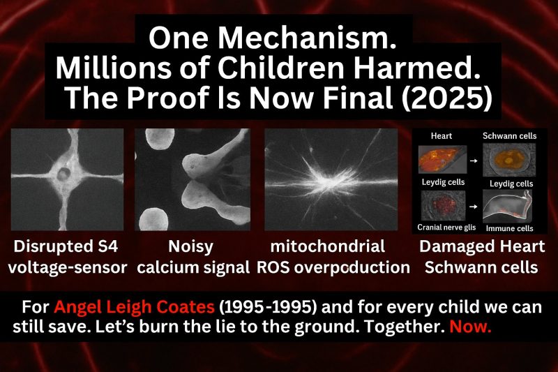
The S4–Mitochondria Rosetta Stone
Why Cancer, Infertility, and Autoimmune Chaos All Point to the Same First Domino
For 30 years we’ve pretended RF radiation is a mystery box: lots of scattered findings, no clear mechanism, so “no proven harm.”
That story is now obsolete.
When you line up the best mechanistic work (Panagopoulos, Jangid, Durdík, etc.), the big animal bioassays (NTP, Ramazzini), and the new WHO-commissioned systematic reviews (SR4A and friends), you do not get noise. You get the same simple chain over and over:
RF/ELF → S4 timing errors in voltage-gated ion channels → distorted Ca²⁺ waveforms → mitochondrial ROS → tissue-specific breakdown.
Once you see that, three “macro-damage” vectors stop looking mysterious:
-
Cancer vector: heart and cranial nerve/glial tissues.
-
Fertility vector: Leydig cells and male germ cells.
-
Autoimmune vector: immune cells decoding Ca²⁺ timing as danger vs tolerance.
This is the Rosetta Stone: one physical entry point (S4 gating in VGICs), one metabolic amplifier (mitochondria), and three downstream systems (heart/brain, testis, immune) that all show convergent damage in the real data.
First domino: S4 and the timing code of life
Every electrically excitable cell in the body – neurons, cardiomyocytes, Leydig cells, T cells – relies on voltage-gated ion channels (VGICs). Each VGIC has:
-
Four homologous domains, each with six transmembrane helices (S1–S6).
-
The S4 helix in each domain studded with positively charged residues. That S4 segment is the voltage sensor. Tiny changes in local electric field make S4 move, which opens or closes the channel.
S4 is where the cell “hears” the outside world as millivolt changes in membrane potential.
Panagopoulos and colleagues showed how polarized, modulated RF/ELF fields can corrupt that hearing. In his ion forced-oscillation mechanism, the RF/ELF field doesn’t have to wrench S4 directly; it only has to shake near-membrane ions:
-
RF/ELF drives forced oscillation of ions in the nanometer-thin aqueous layer around the channel.
-
Those oscillating charges exert strong Coulomb forces on the S4 charges (scaling roughly as 1/r³).
-
The result is irregular, untimely S4 movements – channels opening or closing off-schedule.
For cells whose function is precise timing – heart rhythm, neuronal firing, Ca²⁺ pulses for hormone secretion, Ca²⁺ spikes that drive NFAT / NF-κB in T cells – that is not a small perturbation. It is exactly the kind of timing noise that can derail the whole computation.
That’s your first domino.
Second domino: mitochondrial density as the RF “amplifier”
The next part of the Rosetta Stone is simple:
The more mitochondria a cell has, the bigger the oxidative stress burst when its Ca²⁺ timing is corrupted.
Durdík et al. 2019 gave one of the cleanest proofs of principle. They took umbilical cord blood cells and sorted them along the lineage:
-
Stem / progenitor → more differentiated immune cells.
All populations were pulsed with 2.14 GHz UMTS-like RFR at SAR ≈ 0.2 W/kg. After 1 hour of exposure, they found:
-
ResearchGateROS was increased after 1 h of UMTS exposure (transient – gone by 3 h).
-
Critically: ROS levels rose with the degree of cellular differentiation.
Translation: as cells move from stem → progenitor → mature immune cells, mitochondrial content goes up, and so does the RF-induced ROS burst. That is exactly what you’d expect if mitochondria are the main amplifier of RF timing noise.
You see the same pattern everywhere:
-
Testis, brain, and heart tissues – all mitochondria-dense – are consistently more susceptible to RF-induced oxidative stress and DNA damage than liver, skin, or kidney in comparative animal studies.
-
Reviews on EMF and reproduction highlight mitochondria as both major sources and targets of ROS under RF exposure, especially in germ cells and Leydig cells.
-
In ceLLM language: the “latent space” of damage is not uniform. It is weighted by S4 density and mitochondrial density. Cells that combine both are the hot spots.
The cancer vector: heart and cranial nerve/glial tissues
Look first at where the long-term animal carcinogenicity signal is cleanest.
NTP TR-595 – heart Schwannomas and brain gliomas
In the U.S. National Toxicology Program’s 2-year rat studies (900 MHz GSM/CDMA), male rats showed:
-
Increased malignant Schwannomas of the heart,
-
Increased malignant gliomas of the brain,
with clear exposure–response patterns for several groups. These findings led NTP to classify the evidence for heart Schwannoma as “clear” and for brain glioma as “some” evidence of carcinogenic activity.
Ramazzini Institute – base-station-like exposures
The Ramazzini Institute exposed Sprague Dawley rats from prenatal life to natural death to 1.8 GHz GSM “base-station” signals at whole-body SARs as low as 0.001–0.1 W/kg for 19 h/day. They reported:
-
A statistically significant increase in malignant heart Schwannomas in males at the highest SAR.
-
Elevated malignant glial tumors in the brain.
Different lab, different protocol, different exposure geometry – same two targets: heart Schwann cells and brain glia.
A 2025 WHO-commissioned animal-cancer systematic review, summarized by Melnick, concluded there is high-certainty evidence for heart Schwannomas and moderate-certainty for brain gliomas in RF-exposed rodents.
Mechanistically, that is exactly what the S4–mitochondria Rosetta Stone predicts:
Cardiac conduction fibers and cardiac Schwann cells are rich in Naᵥ, Caᵥ, and Kᵥ channels and sit in mitochondria-dense myocardium that must maintain precise rhythmic firing for a lifetime.
Cranial nerves (e.g., vestibular nerve) and surrounding Schwann/glial cells are likewise VGIC-dense and tightly coupled to mitochondrial energetics.
So when RF/ELF timing noise hits S4 in those circuits, the downstream amplifier (mitochondria) is huge. The signal is chronic oxidative stress and DNA damage in exactly those cell populations – which is what the tumor pattern reflects.
The fertility vector: Leydig and germ cells
Next, take the testis.
Leydig cells as S4-dense mitochondrial nodes
Leydig cells are testosterone factories sitting in the testicular interstitium. They convert LH pulses into mitochondrial steroidogenesis:
-
LH → cAMP/PKA and Ca²⁺ signaling,
-
Ca²⁺ entry through T-type Caᵥ channels and related VGICs,
-
Cholesterol transported into mitochondria (StAR) and converted by CYP11A1, then processed in the ER.
Patch-clamp and molecular studies show Leydig cells express:
-
T-type Ca²⁺ channels (Caᵥ3.x),
-
Voltage-gated K⁺ channels,
-
Ca²⁺-activated K⁺ currents tightly coupled to those Ca²⁺ entries.
So from a physics standpoint, Leydig cells are:
-
VGIC-rich (lots of S4 helices),
-
Mitochondria-dense (steroidogenesis is energy-intensive),
-
Driven by Ca²⁺ timing.
They are a textbook “high S4 + high-mito” node.
Jangid 2025 – non-thermal male reproductive damage
The new Reproductive Toxicology review by Jangid et al. pulls hundreds of animal, in-vitro, and clinical findings together and concludes:
-
RF-EMR triggers oxidative stress in male reproductive cells at non-thermal SARs.
-
Leydig cell mitochondria are highly sensitive to RF exposure.
-
RF-EMR impairs testosterone synthesis and steroidogenesis (StAR, CYP11A1, HSD3β).
-
Sperm count, motility, viability, and morphology decline; DNA fragmentation rises.
-
Testicular architecture and the blood–testis barrier are disrupted.
Everything runs through the same path: redox imbalance + mitochondrial collapse + Ca²⁺ signaling disruption.
SR4A + corrigendum – high-certainty pregnancy-rate reduction
WHO’s SR4A (Cordelli et al.) systematically reviewed 117 animal studies plus in-vitro human sperm work. Following GRADE, they found:
-
A significant, dose-related reduction in pregnancy rate when males were exposed to RF-EMF before mating.
-
Multiple adverse effects on sperm count and motility.
After inconsistencies were found and a corrigendum was issued in 2025, the effect size for pregnancy rate was slightly reduced but still significant, and Melnick reports that the certainty of evidence for pregnancy-rate reduction was upgraded from moderate to high.
So we now have:
-
Mechanistic evidence (Jangid 2025) that Leydig and germ cell mitochondria are damaged and testosterone / spermatogenesis are impaired at non-thermal SARs.
-
Formal WHO-commissioned evidence (SR4A + corrigendum) that male exposure reduces pregnancy rate with high certainty in experimental animals.
Again, that is exactly what you would expect if S4 timing noise + mitochondria is the dominant vulnerability pattern.
The autoimmune vector: immune cells decoding Ca²⁺ timing
The third leg of the Rosetta Stone is the immune system.
Immune cells read danger as Ca²⁺ timing patterns
In T cells and many other immune cells:
-
T-cell receptor (TCR) engagement generates Ca²⁺ oscillations whose frequency and duty cycle encode “activation vs tolerance.”
-
Downstream, NFAT, NF-κB, AP-1, and other transcription factors decode those Ca²⁺ waveforms into gene programs: inflammatory, regulatory, anergic, etc.
-
Those Ca²⁺ waveforms again depend on VGICs (including various Caᵥ channels and CRAC complexes) and are modulated by membrane potential. That means S4 timing errors here become immune decision-making errors.
RF and immune dysregulation: the data
A growing immunology literature shows RF/ELF exposures do not leave the immune system alone:
-
Zhao et al. (2022) exposed rats to 1.5 and 4.3 GHz microwaves, alone and in combination. Single and multi-frequency exposures:
-
Caused pathological changes in thymus and spleen.
-
Decreased white blood cells and lymphocytes at multiple time points.
-
Altered expression of large sets of immune-regulation and metabolism genes, with multi-frequency exposure producing immune-suppressive responses (down-regulating T-cell genes, up-regulating B-cell activation genes).
-
-
Yao et al. (2022) reviewed “The biological effects of electromagnetic exposure on immune cells and potential mechanisms”, concluding that RF/ELF fields can modulate cytokine production, T-cell and B-cell activation, and macrophage function via oxidative stress, Ca²⁺ signaling changes, and membrane receptor alterations.
-
Piszczek et al. (2021) in Environmental Research surveyed “Immunity and electromagnetic fields,” highlighting both immune activation and suppression under RF/ELF, mediated largely by oxidative stress and redox-sensitive signaling.
Add in what radiation biology tells us about oxidative stress, mitochondrial DNA release, and innate immune sensors:
Oxidative stress and mitochondrial damage can release mtDNA into the cytosol, which activates cGAS-STING and NLRP3 inflammasome pathways, driving IL-1, type I interferons, and chronic inflammation.
You already summarized this in your S4→autoimmune tweet card:
-
RF/ELF tweaks S4 gating → altered Ca²⁺ waveforms.
-
T cells and other lymphocytes mis-decode timing: thresholds for activation vs tolerance shift.
-
Phagocytes and other innate cells get the same oxidative push, altering proton conductance and oxidase activity.
-
Mitochondria load up on Ca²⁺, generate ROS, and release mtDNA.
-
cGAS-STING, NLRP3, and related sensors read this as “danger,” not “homeostasis,” pushing interferon and interleukin programs toward chronic activation.
-
Cytokine feedback and redox shifts feed back onto channel expression and kinetics, stabilizing a “trained” inflammatory state with reduced tolerance.
That is exactly how small, persistent timing errors turn into autoimmune-like behavior over years: the immune system repeatedly mis-labels “self” and mundane signals as danger because its timing language has been corrupted upstream at S4.
Pulling it all together: one mechanism, three vectors
When you step back, the puzzle pieces that regulators say “don’t add up” suddenly fit:
Mechanistic physics:
Panagopoulos’ ion forced-oscillation model gives a plausible way for polarized RF/ELF to induce irregular S4 gating in VGICs at realistic field strengths.
Metabolic amplifier:
Durdík 2019 shows a direct scaling of RF-induced ROS with cellular differentiation / mitochondrial load in cord-blood cells at non-thermal SAR (~0.2 W/kg).
Reproductive and neuro studies consistently find higher RF sensitivity in mitochondria-dense tissues, especially testis, brain, and heart.
Cancer vector:
NTP TR-595 and Ramazzini both see heart Schwannomas and brain gliomas in RF-exposed rats, and the WHO animal-cancer SR rates evidence high certainty for heart Schwannoma, moderate for glioma.
Fertility vector:
Jangid 2025 shows Leydig mitochondria + germ cells are hit via oxidative stress, mitochondrial collapse, and hormonal disruption.
WHO SR4A + corrigendum: high-certainty evidence that RF-EMF reduces pregnancy rate in exposed male rodents.
Autoimmune vector:
Immune-focused reviews and experiments (Piszczek 2021, Yao 2022, Zhao 2022) show RF/ELF can shift immune profiles, suppress or mis-activate lymphocytes, and rewrite immune-gene expression via oxidative stress and Ca²⁺/membrane signaling.
Radiation biology shows mtDNA-driven cGAS-STING and NLRP3 activation as canonical routes from oxidative stress to chronic inflammation and autoimmunity.
In other words:
The same RF-induced S4 timing noise and mitochondrial ROS explain:
Why heart and cranial nerves grow Schwannomas and gliomas in NTP/Ramazzini.
Why Leydig/germ cells fail, and pregnancy rate drops with high certainty in male-exposed animals.
Why immune cells mis-decode Ca²⁺ timing and slide toward chronic, mis-targeted inflammation.
There is nothing incoherent here. The “we don’t know how it could work” argument is no longer credible.
The line you can use
If you want one tight line for slides and social:
All the serious RF data are pointing at the same first domino: S4 voltage sensors in ion channels. Polarized RF/ELF adds timing noise at S4, mitochondria amplify it into oxidative stress, and the tissues with the highest S4 and mitochondrial density – heart, brain, Leydig cells, and immune cells – are exactly where we see cancer, infertility, and autoimmune-like damage in the real world.
figure set (heart / nerve / Leydig / immune) in SVG or HTML whenever you’re ready, but conceptually the Rosetta Stone is already there:
S4 + ΔVm as the input.
Mitochondrial ROS as the amplifier.
Cancer, infertility, autoimmune-like inflammation as three facets of the same underlying problem.